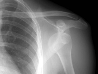
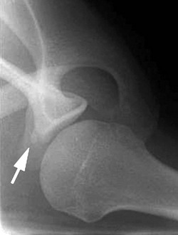
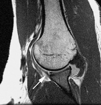
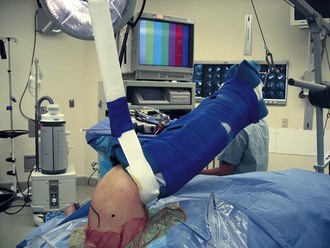
Chapter 4 Recurrent anterior glenohumeral instability is a common sequela of a traumatic glenohumeral dislocation or recurrent subluxation episodes. The major pathoanatomic features of a traumatic dislocation are the capsulolabral avulsion of the inferior glenohumeral ligament (Bankart-Perthes lesion) and capsular redundancy, which typically worsens with repeated injuries.1–5 Once recurrent instability affects activities of daily living or precludes return to sports, operative stabilization is recommended. Although there is still considerable debate about whether to proceed with surgery after the first traumatic dislocation, the orthopedic literature supports early acute arthroscopic stabilization after a traumatic dislocation in a select group of young athletes who are at high risk for repeated shoulder injuries.6–11 However, patient issues such as player position, time remaining in a season, and time available for rehabilitation may affect the decision of when to undergo surgery. Immediate stabilization is recommended in athletes who participate in activities or sports in which a subsequent dislocation could be life-threatening (e.g., parachuting or rock climbing). Arthroscopic shoulder stabilization offers a number of advantages over traditional open repairs. These include smaller incisions, less muscle dissection, less postoperative pain, and better visualization of the entire glenohumeral joint.12 The first arthroscopic Bankart repairs were performed by transglenoid suture fixation. Sutures were passed across the glenoid and tied over the posterior fascia. As bioabsorbable polymers were introduced for use in the shoulder, soft tissue fixation with tacks became popular. The tacks were inserted arthroscopically over a guidewire to ensure correct placement. Tacks have been shown to have limited pull-out strength and have, for the most part, been abandoned for shoulder stabilization. The success of metallic suture anchors in open shoulder surgery led to the development of arthroscopic deployment techniques. Development of longer-lasting polymers and high-strength suture made bioabsorbable anchors the most popular choice for soft tissue repair. The goals of any suture anchor are to repair soft tissue to bone and to be able to withstand the forces required for rehabilitation until the normal bone-tissue interface is restored. The focus of this chapter is on the technique of arthroscopic anterior shoulder stabilization with use of suture anchors. Recurrent instability causes apprehension when the shoulder is in the abducted, externally rotated position. Relief of the apprehension with posteriorly directed pressure on the proximal humerus, the relocation sign, is often present. Glenohumeral patholaxity can be assessed and graded in comparison with the contralateral side. This examination should be repeated with use of anesthesia, when a better comparison with the normal side can be achieved. The examination under anesthesia includes the supine load-shift test with the arm abducted at 70 to 90 degrees to document and to quantify the degree of anterior instability of the glenohumeral joint compared with the contralateral side. This test can be performed with the patient supine or sitting upright. Another sign of anterior instability is the scapular protraction sign described by Bottoni.13 In the seated position, as the abducted arm is slowly externally rotated, the patient may involuntarily protract the scapula to keep the glenoid articulating with the humeral head. This will be identified as a winging appearance of the scapula with increasing external rotation of the shoulder. • A standard anteroposterior view with the arm in slight internal rotation is used to identify fractures of the greater tuberosity and Hill-Sachs lesions (Fig. 4-1). • The trans-scapular Y view can assist with the direction of dislocation before reduction and confirm successful reduction. • The West Point axillary view can be used to assess glenoid rim fractures (bony Bankart lesion; Fig. 4-2). • Computed tomography (CT) is occasionally used to assess the extent of bone injuries of the humerus or glenoid. In addition, CT can be used to evaluate the glenoid version. • Magnetic resonance imaging is the gold standard for evaluation of intraarticular pathoanatomy. For evaluation of recurrent instability, we prefer magnetic resonance arthrography (MRA) because the addition of intraarticular gadolinium distends the shoulder joint and improves the visualization of the pathoanatomy (Fig. 4-3). After an acute dislocation or subluxation, the hemarthrosis serves to distend the joint and obviates the need for contrast agent. The primary operative indication for a stabilization procedure is shoulder instability that interferes with activities of daily living or recreational sports. Recurrent dislocation or subluxation episodes can result in additional chondral or osteochondral damage. Contraindications to surgery include habitual or voluntary dislocators and unwillingness or inability of a patient to comply with the obligatory postoperative restrictions and rehabilitation program. Bone defects such as a large bony Bankart or an engaging Hill-Sachs lesion are best treated with open surgical techniques. A relative contraindication to arthroscopic stabilization includes recurrent instability in athletes involved in contact sports.14 Anterior stabilization is typically performed with the patient under general anesthesia. An adjunctive regional (interscalene) block may be performed to provide postoperative analgesia. Positioning of the patient is based on the surgeon’s preference. Many surgeons believe that the lateral decubitus position allows better visualization and ease of instrumentation with a shoulder distraction system (STaR Sleeve and 3-Point Shoulder Distraction System, Arthrex, Naples, FL; Fig. 4-4). However, the beach chair position may allow greater control of the entire arm, especially internal and external rotation. This position also facilitates easier conversion to a traditional open approach.
Suture Anchor Fixation for Anterior Shoulder Instability
Preoperative Considerations
Physical Examination
Imaging
Other Modalities
Indications and Contraindications
Surgical Technique
Related posts:
 Knot-Tying and Suture-Passing Techniques
Open Repair of Posterior Shoulder Instability
Central Quadriceps Free Tendon Harvest for Anterior Cruciate Ligament Reconstruction
Patient Positioning, Portal Placement, and Normal Arthroscopic Anatomy
Arthroscopic and Open Decompression of the Suprascapular Nerve
Allografts for Anterior Cruciate Ligament Reconstruction
Knot-Tying and Suture-Passing Techniques
Open Repair of Posterior Shoulder Instability
Central Quadriceps Free Tendon Harvest for Anterior Cruciate Ligament Reconstruction
Patient Positioning, Portal Placement, and Normal Arthroscopic Anatomy
Arthroscopic and Open Decompression of the Suprascapular Nerve
Allografts for Anterior Cruciate Ligament Reconstruction
![]()
Stay updated, free articles. Join our Telegram channel

Full access? Get Clinical Tree


Suture Anchor Fixation for Anterior Shoulder Instability