Surgical Management of Malignant Bone Tumors Around the Knee

Chapter 149 Surgical Management of Malignant Bone Tumors Around the Knee


The surgical management of malignant tumors about the knee requires a basic understanding of tumor biology, knowledge of common tumors, and their clinical and radiographic behavior. To treat malignant tumors successfully, it is essential to understand general principles of sarcoma surgery, including staging of a bone tumor, appropriate biopsy techniques, patient selection for limb salvage surgery versus amputation, and methods of resection and reconstruction. These topics will be discussed in this chapter. Initial workup, including staging and biopsy of a bone tumor, are covered elsewhere in this text. Reconstructive options that will be reviewed include arthrodesis, allograft reconstruction, and rotationplasty. Reconstruction with a megaprosthesis or allograft-prosthetic composite are discussed elsewhere. Common malignant bone tumors around the knee include osteosarcoma, chondrosarcoma, Ewing’s sarcoma, and lymphoma of bone, and these will also be reviewed.


Tumor Biology


Primary malignant tumors have the capacity to metastasize to distant sites. They also are locally aggressive, destroy bone, and involve adjacent soft tissue structures. Malignant tumors are typically subclassified as low, intermediate, or high grade based on the histologic grade. Grade is a feature of the amount of cellular activity, cellular pleomorphism, mitotic activity, and tumor necrosis. It is reflective of the tumor’s aggressiveness.


Malignant bone tumors spread locally by centrifugal tumor expansion. As the tumor cells divide and the mass grows, normal tissue is compressed. Microscopic pseudopods of tumor invade normal surrounding tissue. In an effort to control the tumor, an inflammatory response is established adjacent to the tumor. This reactive zone consists of inflammatory cells, edematous tissue, and neovascularity feeding the advancing tumor. The compressed normal tissue produces a pseudocapsule about the mass. Thus, the surrounding soft tissue around the malignant tumor is slowly invaded by satellites of advancing tumor. To remove the tumor completely, the resection must be well beyond the reactive zone and into normal adjacent soft tissue of bone, thus avoiding leaving satellites behind.12 Unfortunately, current imaging modalities such as magnetic resonance imaging (MRI), positron emission tomography (PET) scan, and bone scanning cannot identify microscopic tumor extension adequately. Often, the edematous tissue can be well identified, but this really provides no clear information regarding the presence or absence of microscopic tumor satellites.


This aggressive local spread of tumor is frequently halted by anatomic barriers, such as cortical bone, periosteum, cartilage, synovium, and fascia. These barriers are relative and do not offer an absolute stopping point for tumor growth. They can be breached with tumor extension beyond their confines. However, they do serve to force tumor growth into a longitudinal pattern. Tumors can extend for great distances along bone and soft tissue planes and attain a large bulky size before being detected. Tumors that remain within the confined bone or soft tissue compartment are termed intracompartmental.2 Tumors that extend out of the compartment of origin and involve adjacent structures are termed extracompartmental. As the tumor spreads, it frequently displaces rather than encases neurovascular structures. Occasionally, these structures can be circumferentially engulfed by tumor but tend to remain patent and functional unless significantly compressed by a large mass. Destruction of bone and presence of a soft tissue mass can lead to pain, the most common presenting symptom of malignant tumors of bone. The subcutaneous nature of the distal femur and proximal tibia lead to early detection of a soft tissue mass associated with a bone tumor.


Principles of Surgical Management


On completion of appropriate staging and the determination of an accurate diagnosis, attention is now turned toward treatment. Cure of malignant tumors requires local control of tumor as well as control of systemic disease. Surgical management of malignant bone tumors is often the foundation for local control. The principles of surgical management are outlined here. Adjuvant treatments for common malignant tumors are found elsewhere in this text in discussions of each of the common bone malignancies.


Surgical Margins


In collecting, analyzing, and reporting surgical data, it is imperative to use a language that is common among surgeons as to which surgical procedure has been performed. The relationship between the surgical plane of resection of the tumor and surrounding tissue is known as the surgical margin. Enneking and Shirley3 have established a nomenclature to describe oncologic surgical procedures in terms of four surgical margins. The margin can be achieved by amputation or a limb salvage procedure.


Intralesional margins are achieved if the surgical plane of dissection is through the tumor, leaving behind gross residual disease. This typically occurs when a biopsy is done but also with curettage or a debulking procedure. Tumor is obviously left behind and the procedure is not curative for malignant bone tumors. This margin is appropriate only in the palliative setting or for very low-grade lesions amenable to local control with curettage alone.


Marginal margins are achieved when the plane of dissection is the pseudocapsule itself. The main body of the tumor easily peels away from the surrounding pseudocapsule. This leads the surgeon to perform a shell-out procedure inadvertently. Failure to recognize that histologically, there is a real potential to have satellites of tumor within and beyond the pseudocapsule, which results in lack of adequate margin. Several studies have demonstrated the high rate of tumor cells in this reactive zone after a shell-out or gross total resection. Even after adjuvant therapies, local recurrence is high with a marginal margin. This is why tumor bed re-excisions are often recommended after gross total resections or shell-out procedures are performed. In tumors treated preoperatively with chemotherapy or radiation, a marginal margin can be accepted only adjacent to vital structures such as a neurovascular bundle. In this situation, the preoperative treatment would have (hopefully) sterilized any microscopic satellite lesions in these areas.


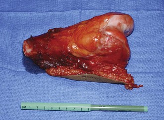
Wide margins are the most commonly achieved margins when dealing with bone malignancies. The resection plane is beyond the pseudocapsule and reactive zone. It results in a cuff of normal tissue surrounding the tumor. The specimen contains the biopsy tract (skin and underlying soft tissue), body of the tumor, pseudocapsule, reactive zone, and a cuff of normal tissue. The thickness of that cuff is subject to study and debate. It must be realized that its thickness is not as important as the concept that the cuff is wide enough to be beyond any satellite or skip lesions that may be present. A wide margin is the goal of surgical treatment in bone and soft tissue sarcomas to minimize the risk of local tumor recurrence12 (Fig. 149-1).


Radical margins are achieved when the involved bone and soft tissue component are resected in their entirety. An extracompartmental resection is thus achieved. When managing tumors that are extracompartmental, the entire involved bone and involved adjacent musculature must be excised to be considered a radial resection. Radical margins are typically unnecessary for bone sarcomas.


Principles of Tumor Resection


Once a patient is considered a candidate for limb salvage, careful preoperative planning of the resection and reconstruction is essential to achieving a successful outcome. Review of initial and interval imaging studies during the course of treatments allows the surgeon to plan the definitive surgery. Bone resection is determined by initial tumor imaging and is typically done with a 3- to 5-cm resection of bone to achieve a wide surgical margin. In some cases, this margin may be as close as 1 cm. The subchondral bone provides an excellent barrier to tumor extension. Even when tumor extends into the epiphyseal bone, it is rare for tumor to extend into the joint. This allows for tumor resection of the proximal tibia or distal femur in an intra-articular fashion. Tumors that involve the proximal tibia often require removal of the proximal fibula through the proximal tibia–fibula joint if the joint is in danger of tumor penetration or in close proximity to tumor. Tumors of the proximal fibula are usually treated with resection of the fibula alone but may require removal of the proximal tibia/fibula joint in an extra-articular fashion, removing a small portion of the adjacent tibia. Large tumors or tumors with an intra-articular extent require resection in an extra-articular fashion. If there has been tumor extension into the knee joint or contamination from a previous ill-planned biopsy, the joint must be removed en bloc by resection beyond the capsular attachments at the proximal tibial and distal femoral levels. The patella and extensor mechanisms are resected along with the joint. The resection must include any skip metastases in the bone or soft tissues.


Special consideration is also given for the skeletally immature child who has significant growth remaining. Resection of the distal femoral and proximal tibial physes can result in a significant limb length inequality. Special reconstruction options must be considered for these growing children. Amputation, rotationplasty, and an expandable prosthesis are reconstructive options for this group of patients. Resection level may be influenced by the desire to save the physis, but always the best oncologic margin is what determines the resection level.


Once the bone margins are determined, consideration is given to the soft tissue resection. Most tumors of the distal femur are adequately covered by deep layers of soft tissue. Frequently, an adequate amount of quadriceps mechanism may be spared to provide an appropriate functional result. Usually, only a small portion of the vastus medialis oblique or vastus lateralis is removed with resection of the biopsy track. The remainder of the quadriceps, patella, and patellar tendon can be spared. Typically, the deep soft tissue margin (providing coverage over the tumor) is provided by the vastus intermedius. In large bulky tumors, more extensive quadriceps resection may be required. This may have an effect on function and should be considered when deciding on amputation versus limb salvage. The neurovascular bundle may be displaced by the sarcoma but is usually not encased by tumor and can be spared. The cruciate ligaments, collateral insertions, and gastrocnemius insertions are all removed off the distal femur, requiring a constrained design if arthroplasty is the method of skeletal reconstruction. The adductors are divided as well.


The pes anserine and hamstring insertions on the tibia and fibula are usually left intact when performing distal femoral resections. Resection of ligamentous insertions is always done with a short cuff of tissue to ensure adequate margins. Typically, the resection is performed by first dissecting through soft tissues, which allows the biopsy tract to be removed with the specimen, performing the arthrotomy, continuing with the soft tissue dissection, and then osteotomizing the femur. Often, the posterior soft tissues are more easily approached after the femoral osteotomy has been performed. The specimen is removed and inspected by the surgeon and pathologist for margins. Attention is then turned toward reconstruction.


On the tibial side, tumors frequently involve the tibial tubercle. This requires division of the patella tendon, which must be reconstructed to provide an adequate extensor mechanism. The pes anserine and hamstring insertions are divided near the bone with a cuff of normal tissue. On the femoral side, the gastrocnemius and adductor insertions are spared. The cruciate and collateral attachments are removed from the proximal tibia as well when resecting the proximal tibial lesion.

Stay updated, free articles. Join our Telegram channel

Aug 26, 2016 | Posted by in ORTHOPEDIC | Comments Off on Surgical Management of Malignant Bone Tumors Around the Knee

Full access? Get Clinical Tree

Get Clinical Tree app for offline access