Chapter 149 Surgical Management of Malignant Bone Tumors Around the Knee
Tumor Biology
Malignant bone tumors spread locally by centrifugal tumor expansion. As the tumor cells divide and the mass grows, normal tissue is compressed. Microscopic pseudopods of tumor invade normal surrounding tissue. In an effort to control the tumor, an inflammatory response is established adjacent to the tumor. This reactive zone consists of inflammatory cells, edematous tissue, and neovascularity feeding the advancing tumor. The compressed normal tissue produces a pseudocapsule about the mass. Thus, the surrounding soft tissue around the malignant tumor is slowly invaded by satellites of advancing tumor. To remove the tumor completely, the resection must be well beyond the reactive zone and into normal adjacent soft tissue of bone, thus avoiding leaving satellites behind.12 Unfortunately, current imaging modalities such as magnetic resonance imaging (MRI), positron emission tomography (PET) scan, and bone scanning cannot identify microscopic tumor extension adequately. Often, the edematous tissue can be well identified, but this really provides no clear information regarding the presence or absence of microscopic tumor satellites.
This aggressive local spread of tumor is frequently halted by anatomic barriers, such as cortical bone, periosteum, cartilage, synovium, and fascia. These barriers are relative and do not offer an absolute stopping point for tumor growth. They can be breached with tumor extension beyond their confines. However, they do serve to force tumor growth into a longitudinal pattern. Tumors can extend for great distances along bone and soft tissue planes and attain a large bulky size before being detected. Tumors that remain within the confined bone or soft tissue compartment are termed intracompartmental.2 Tumors that extend out of the compartment of origin and involve adjacent structures are termed extracompartmental. As the tumor spreads, it frequently displaces rather than encases neurovascular structures. Occasionally, these structures can be circumferentially engulfed by tumor but tend to remain patent and functional unless significantly compressed by a large mass. Destruction of bone and presence of a soft tissue mass can lead to pain, the most common presenting symptom of malignant tumors of bone. The subcutaneous nature of the distal femur and proximal tibia lead to early detection of a soft tissue mass associated with a bone tumor.
Principles of Surgical Management
Surgical Margins
In collecting, analyzing, and reporting surgical data, it is imperative to use a language that is common among surgeons as to which surgical procedure has been performed. The relationship between the surgical plane of resection of the tumor and surrounding tissue is known as the surgical margin. Enneking and Shirley3 have established a nomenclature to describe oncologic surgical procedures in terms of four surgical margins. The margin can be achieved by amputation or a limb salvage procedure.
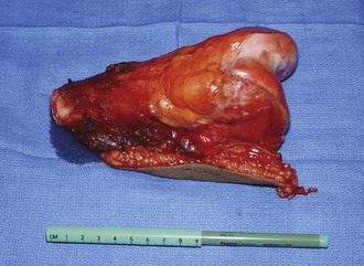
Wide margins are the most commonly achieved margins when dealing with bone malignancies. The resection plane is beyond the pseudocapsule and reactive zone. It results in a cuff of normal tissue surrounding the tumor. The specimen contains the biopsy tract (skin and underlying soft tissue), body of the tumor, pseudocapsule, reactive zone, and a cuff of normal tissue. The thickness of that cuff is subject to study and debate. It must be realized that its thickness is not as important as the concept that the cuff is wide enough to be beyond any satellite or skip lesions that may be present. A wide margin is the goal of surgical treatment in bone and soft tissue sarcomas to minimize the risk of local tumor recurrence12 (Fig. 149-1).
Stay updated, free articles. Join our Telegram channel

Full access? Get Clinical Tree


