Fig. 20.1
Performing a cutback. Surfer: Kenneth Taylor. El Salvador
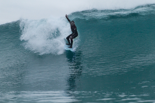
Fig. 20.2
Dropping into an overhead wave. Surfer: Kenneth Taylor. Spot: Blacks Beach, San Diego (CA)
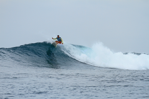
Fig. 20.3
Top turn. Surfer: Kenneth Taylor. Indonesia
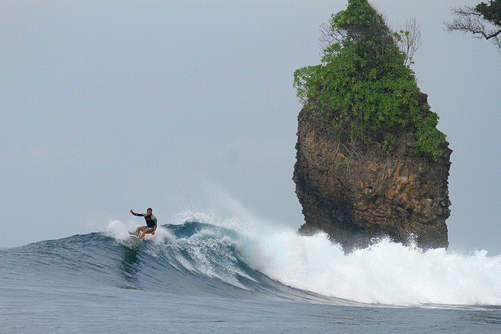
Fig. 20.4
Island surf. Surfer: Kenneth Taylor. Indonesia
Stand-up paddle surfing is growing in popularity. It is a variant of surfing, in which the surfer paddles to move through the water, and glides or rides waves (Fig. 20.5) while standing on a surfboard.
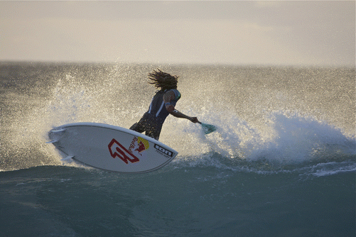
Fig. 20.5
SUPing in Cape Verde. Surfer: Airton Cozzolino (Photo courtesy of Claudio Marosa)
With the advent of newer technological means of creating rideable waves in pools and indoor arenas, surfing has expanded from coastal and great lake communities to inland areas and even cruise ships.
Surfing is an intermittent sport with paddling accounting for 50 % of the activity, limited motion/waiting for suitable waves for 40 %, and 5–10 % spent actually riding the wave. Surfers possess a high level of aerobic fitness and peak VO2 values are comparable to other upper-body endurance-based athletes. There are cyclical bouts of low-intensity activity soliciting aerobic metabolism intermixed with high-intensity exercise utilizing both aerobic and anaerobic metabolism [2]. As a highly active water sport, those participating in surfing are prone to a unique constellation of acute and chronic conditions which physicians caring for these patients should be aware. This chapter will review some of the more frequent illnesses and injuries seen in these athletes with a discussion of relevant preventive strategies.
20.2 Trauma
Surfboard riders are prone to a multitude of acute and chronic conditions (Table 20.1).
Table 20.1
Summary of common injuries seen in surfboard riders
Acute injuries | Chronic injuries |
|---|---|
Head lacerations | Repetitive motion injuries to back, shoulders, knees, neck |
Lower extremity lacerations | Auditory exostoses |
Lower extremity sprains, especially knee | Otitis externa |
Contusions | Pterygium |
Concussion | Recurrent cellulitis |
Marine envenomation | |
Tympanic membrane rupture |
Several authors have presented retrospective studies on the frequency and type of injuries incurred by surfers. Lowden et al. published one of the first studies on the prevalence of injuries among 346 Australian surfers which required either medical attention or days lost from surfing [3]. They reported that lacerations were most frequent, representing 41 % of all injuries. Most lacerations were to the head, mainly to the skull, with lower extremity lacerations next most frequent. The second most common injury type was dislocations, sprains, and strains which represented 35 % of all injuries. Other injuries included skull and body fractures, contusions, and tympanic membrane perforations. Overall, 3.5 injuries per 1000 surfing days were reported in their study.
In 2002, Nathanson published a larger study of 1348 surfboard riders [1]. They also found lacerations to be the most common injury, but with head and neck lacerations to be equally as prevalent as lower extremity ones. Contusions, sprains, and fractures were also seen. The majority of contusions affected the trunk and lower extremity. Sprains were most reported in the lower extremity, with knee injuries most often seen. The majority of fractures reported by Nathanson et al. were to the head and neck. Of note, both Lowden and Nathanson found a higher level of injury severity in more advanced surfers, who often engage larger waves in more extreme conditions (Fig. 20.6).
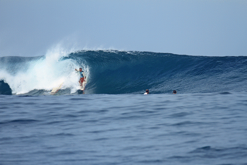
Fig. 20.6
Barreling wave, nearby surfer bails his board (yellow board) which gets broken in half by the wave
The large majority of injuries reported in both studies were from contact with a surfboard, usually the rider’s own board. The rail and fins of the board accounted for most of the injuries seen. The seafloor is another common source for injury, responsible for 17 % of injuries in the Nathanson study. Surfing over coral reefs versus sand is a significant risk for sea floor injuries [1] (Fig. 20.7).
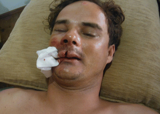
Fig. 20.7
Facial lacerations and contusions from contact with reef
Additional risks involved with surfing include injury leading to either drowning or hypothermia should the surfer not be able to safely return to shore. Lacerations have the added potential of becoming infected with seaborne organisms and should be treated for Vibrio and Pseudomonas, along with the more common Staphylococcus and Streptococcus.
Nathanson published another study in 2007 on injuries during 16,657 surfer heats observed over 32 surfing competitions worldwide from 1999 to 2005 [4]. They found an injury rate of 13 per 1000 h of competitive surfing. Professional competitions had higher rates of injury in comparison to amateur competitions; however, professional contests were held in larger surf, were longer in duration, and occurred more often over a hard bottom, as opposed to sand. Sprains and strains were most commonly seen, followed by lacerations, contusions, and fractures. The lower extremity was the most injured location, with knee sprain/strain being the most common injury pattern overall. The majority of injuries were due to impact with the surfboard, and most injuries occurred during an unsuccessful takeoff, followed by turning maneuvers.
Preventive safety measures can help reduce the frequency and severity of injury. Surfing helmets are available and their widespread use could help reduce the number of head lacerations and fractures [5]. Nathanson, however, reported that only 8 % of surfers in their survey used helmets [1]. Rubber guards on the board’s nose and soft-edged or rubber-guarded fins would also help reduce the number of lacerations and are thought not to alter surfboard dynamics to any appreciable degree. Their use as reported by Nathanson is limited, with 40 % of surfers reported using nose guards and only 5 % of surfers having soft-edged fins [1]. Protective eyewear specifically designed for surfing is available from several manufacturers and may afford both protection from UV rays and orbital trauma.
The use of a board leash in preventing injury is controversial. Leashes do seem to have reduced the number of accidents involving loose boards hitting other surfers. Leashes also provide the downed surfer with access to a floatation device in the event of serious injury. But by keeping the board in close proximity to the surfer, leashes may increase the risk of board-induced injury. In addition, board recoil from the leash is another mechanism for injury to the surfer. Two articles of surfboard-related ocular trauma implicate the board nose as the common mechanism of injury, with Kim et al., implicating leash recoil as one causative factor [6, 7]. Leashes are sold in varying lengths. Longer leashes may decrease recoil injury, with the consequence of increased risk of injury to others.
A recent addition to potential injury associated with surfing has been coined surfer’s myelopathy by Thompson et al., who reported nine cases occurring between 1998 and 2003 in Hawaii [8]. Since then, several case reports and case series have been published, describing very characteristic clinical presentations and features among patients with surfer’s myelopathy [9–18]. It seems to affect first-time surfers or those learning the sport. It appears to be a nontraumatic paraparesis/paraplegia that may be associated with hyperextension of the lower thoracic, upper lumbar segments in an untrained surfer. No injury was noted during surfing, but most patients complained of mild mid- to low back pain, lower extremity weakness, and urinary retention. A retrospective review of MR findings in 23 cases of surfer’s myelopathy by Nakamoto et al. demonstrated central T2 hyperintense signal abnormalities in the spinal cord extending from the midthoracic region to the conus in all cases. These findings were associated with cord expansion and conus enlargement, all seen within 24 h of symptoms onset. However, MR findings did not correlate with symptom severity or clinical improvement [19]. Although most patients achieved significant improvement or resolution, several cases of complete paraplegia have been noted. The pathophysiology is possibly due to a secondary ischemic event from hyperextension and spinal cord traction, resulting in spinal cord injury and the abnormal signal findings of the lower thoracic/upper lumbar segments of the spinal cord seen on MRI. While not enough is known of this disorder to make definitive recommendations on prevention, swimming and surfboard paddling for endurance and strength gains as well as flexibility training may reduce the incidence of this malady, particularly since it does not appear to affect experienced surfers.
Surfing like most other sports is not immune to overuse injuries. The overhead nature of paddling is similar to swimming. Shoulder impingement syndrome, acromioclavicular arthrosis, and rotator cuff strains are common to surfers. Treatment is similar to that seen in other sports and includes activity modification, rotator cuff/periscapular strengthening, injections, and arthroscopic surgery for refractory cases or complete cuff tears. Prevention is geared toward a strengthening and consistent paddling regimen to maintain adequate strength and fitness [2].
Neck and lower back pain are common complaints of surfers. Degenerative disk and joint disease account for a fair amount of these complaints, particularly among aging surfers. Surfing has been implicated in a number of cases of spondylolysis and spondylolisthesis due to its repetitive hyperextension of the lumbar spine [2].
Spine trauma in the cervical spine is of particular concern because of the potential for central cord syndrome (CCS). Over 50 % of these injuries occur in older surfers particularly those with preexisting spondylosis [20]. The mechanism of injury was similar with nearly all of the injuries caused by hitting the ocean floor (75 %) resulting in neck hyperextension [21]. Injuries usually occur in the lower cervical spine and can result in burst fractures and complete spinal cord injuries.
20.3 Illnesses
20.3.1 Marine Envenomation
Interaction with other sea life is an inevitable part of ocean sports. Marine animals caused 3 % of reported injuries in the study by Nathanson. The more frequently encountered life forms which cause harm to surfers are the free-floating coelenterates, stingrays, and coral reefs. While clinical presentation may vary, certain general treatment principles apply to any type of marine envenomation.
First, most marine envenomations become infected [21]. Common organisms include Staphylococcus, Streptococcus, and Vibrio species. Special culture medium is needed for growing marine organisms, and the lab should be alerted if there are concerns for these. Broad-spectrum empiric coverage is warranted, and either third-generation cephalosporins or fluoroquinolones are good choices to treat Vibrio species. Lacerations should be allowed to heal secondarily or, if necessary, by delayed primary closure. Second, retained foreign bodies should be considered in most envenomations. Depending on the mechanism of injury and level of clinical suspicion, investigation of a retained foreign body can be done through wound exploration or appropriate radiographs. Tetanus prophylaxis should be given if the patient’s immunity is not up-to-date.
20.3.1.1 Coelenterates
Coelenterates are invertebrate animals and can be either free floating or sessile. Surfers are more apt to encounter free-floating coelenterates such as the true jellyfish, box jellyfish, and Portuguese man-o-war. These animals have a main body and multiple dangling tentacles with numerous venom-filled cells called nematocysts. Nematocysts inject their toxins subcutaneously in response to either chemical or mechanical stimuli. Local symptoms of nematocyst envenomation include burning pain, erythema, edema, urticaria, and bullae formation, all which may lead to skin necrosis (Fig. 20.8). Systemic effects of the toxin can damage respiratory, cardiovascular, gastrointestinal, renal, musculoskeletal, neurologic, and ocular organs.
Fig. 20.8
Jellyfish stings
Initial treatment involves preventing further toxin release by removing any remaining tentacles or other animal parts. Larger animal parts can be carefully removed with forceps or gloves. No consensus exists as to the best method of inactivating nematocysts and reducing pain. Various treatments recommended by published and anecdotal sources include cold packs, heat application, or irrigation with a multitude of liquids. Study results are conflicting due to the variety of study designs and jellyfish species used (Table 20.2).
Table 20.2
Summary of study results conducted to evaluate different treatment strategies to reduce pain associated with jellyfish stings
Study | Nematocyst type | Treatment | Findings |
|---|---|---|---|
Hartwick et al. [22 ] | Chironex (indo-pacific box jellyfish/sea wasp) | Methylated spirits | Released toxin |
Ethanol | Released toxin | ||
Urine | Released toxin | ||
Acetic acid | Prevented toxin release | ||
Vinegar | Prevented toxin release | ||
Burnett et al. [23 ] | Chrysaora (sea nettle) | Vinegar | Caused nematocyst rupture |
Chrysaora | Baking soda | Prevented toxin release | |
Physalia (bluebottle/Portuguese man-o-war)
Related posts:Stay updated, free articles. Join our Telegram channel
Full access? Get Clinical Tree
 Get Clinical Tree app for offline access

|
