Supramalleolar Osteotomy
Shannon M. Rush
John M. Schuberth
Realignment osteotomy of the distal tibia is a valuable surgical procedure for the treatment of distal tibial malalignment resulting from posttraumatic malunion, physeal disturbances, congenital and metabolic diseases, and degenerative arthritis (1,2,3,4,5,6,7,8,9,10 and 11). Juxta-articular malalignment of the distal tibia often results in hindfoot and forefoot compensation, which creates predictable patterns of joint degeneration and pain. However, small degrees of malalignment are generally well tolerated if there is no associated stiffness or arthritis in the hindfoot (12,13). The rationale for supramalleolar osteotomy (SMO) is to preserve the joints of the foot and ankle from articular degeneration and biomechanical dysfunction by restoring the joint orientation and axial alignment of the limb. Furthermore, the realignment maneuver may be helpful in alleviating symptoms secondary to degenerative changes by readjusting the forces of weight-bearing on the articular surfaces. Lastly, these procedures may serve a secondary purpose of improving postural or suprastructural symptoms of the hip, knee, and spine during gait. The absolute magnitude of necessary deformity to indicate an SMO is not clear and must be taken in the context of clinical condition.
PATHOMECHANICS
Malposition of the distal tibiofibular complex can have significant functional and biomechanical consequences. These problems include accelerated focal wear of the articular surfaces of the foot and ankle and abnormal gait patterns (14,15). The degree to which the deformity influences clinical symptoms and function depends on several factors. The degree of motion in the subtalar and midtarsal joint, size of individual, severity of index injury, intra-articular fracture patterns, articular incongruity, and age all contribute to the effect the deformity will have clinically. The available amount of motion in the subtalar joint is not precisely understood and can vary considerably between patients (16,17). Paley (18) believes 30 degrees of ankle valgus and 15 degrees of ankle varus can be compensated for with a normal functioning subtalar joint. Distal tibial valgus deformity is better compensated than distal tibial varus deformity because twice as much inversion exists in the subtalar joint than eversion. When the distal tibial deformity exceeds the available frontal plane compensation in the subtalar joint, several clinical scenarios develop. The amount of ankle varus deformity exceeds the available subtalar eversion, and residual hindfoot varus deformity results. This condition creates additional forefoot pronation to maintain a plantar grade foot. The opposite occurs when a distal tibial valgus deformity is incompletely compensated with subtalar joint inversion. Complicating this condition is the progressive development of degenerative stiffness in the subtalar joint and inability to compensate for distal tibial deformity. Frequently, an associated injury to the subtalar joint renders normal compensatory motion inadequate to adjust to a supramalleolar deformity.
Tarr (14) showed distal tibial deformity significantly altered the contact location, shape, and magnitude of the tibiotalar contact pressures. Sagittal plane malalignment created the most significant alterations in contact characteristics. Recurvatum (shear deformity) and procurvatum (impingement deformity) of 15 degrees resulted in changes in contact biomechanics of greater than 40%. These cadaver studies showed how deformity near the ankle joint can focus contact pressure to small areas of the articular surface. This could explain why deformity is initially well tolerated in the distal tibia and often presents only later when painful degenerative articular wear patterns begin in the ankle and subtalar joint. These cadaver studies are difficult to extend into the clinical situation but do correlate with the clinical patterns of articular degeneration observed clinically. Steffensmeier (19) demonstrated the focal areas of the talar dome could be off-loaded with shift in center of pressure of 1 and 1.58 mm with lateral and medial displacement osteotomy of 1 cm, respectively. This shift in ground reactive force (GRF) demonstrates the ability to manipulate the GRF acting on the hindfoot and ankle with corrective osteotomy. The shift in GRF demonstrates the ability to manipulate the GRF acting on the hindfoot and ankle with corrective osteotomy. Normally, GRF passes through the heel and lateral aspect of the ankle joint, creating a valgus torque in the hindfoot (20). The lateral position of the calcaneal axis with respect to the tibial axis explains this mechanical principle. Additionally, abnormal lateral translation (>1 cm) of the calcaneus can cause detrimental hindfoot valgus forces. Often, the surgeon encounters a dysfunctional flatfoot with hindfoot valgus associated with ankle valgus deformity. This poorly compensated ankle and hindfoot valgus will predictably lead to posterior tibial tendon dysfunction and deltoid ligament failure over time. Understanding the influence of malalignment of the distal tibia and hindfoot is critical to understanding the importance of realignment osteotomy. The foot and ankle surgeon can use these techniques to restore axis alignment and joint orientation and secondarily off-load focal degenerative areas of the ankle. The clinical consequence of realignment osteotomy in preserving the tibiotalar joint in the long term is not well documented, although skeletal malalignment must be corrected prior to arthrodesis or implant arthroplasty.
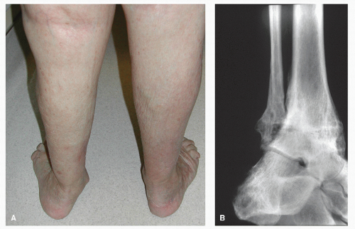
Malpositioned ankle arthrodeses can lead to significant degenerative changes in the subtalar and midtarsal joints with alteration in the GRFs generated in the limb. Fusing the joint with the talus poorly aligned with the axis of the leg will lead to gait problems and forefoot overload. It is preferable in most cases to translate the talus slightly posterior to the tibial axis to alleviate these problems. In addition, sagittal plane malalignment can lead to degeneration of the midtarsal joint and generate recurvatum thrust on the knee. Corrective osteotomy for ankle malunion after arthrodesis is directed at restoring a plantar grade foot and realigning the tibiocalcaneal axis (Fig. 114.1). Growth plate disturbances from fracture or
infection can result in significant secondary deformity. Often, limb length inequality is an additional consideration in these cases, and planning must take excessive shortening into account. McNicol et al (5) and Scheffer and Peterson (6) demonstrated the use of SMO for congenital deformity in children. McNicol used SMO derotational osteotomy in children with complex equinovarus deformity and external tibial torsion. Scheffer described an opening wedge osteotomy (OWO) to correct deformity and restore length. Best (20) described a small series of four patients who had five opening wedge osteotomies with a Puddu plate (Arthex Inc, Naples FL). Autogenous bone was used in all cases, and all osteotomies had healed by 3 months.
infection can result in significant secondary deformity. Often, limb length inequality is an additional consideration in these cases, and planning must take excessive shortening into account. McNicol et al (5) and Scheffer and Peterson (6) demonstrated the use of SMO for congenital deformity in children. McNicol used SMO derotational osteotomy in children with complex equinovarus deformity and external tibial torsion. Scheffer described an opening wedge osteotomy (OWO) to correct deformity and restore length. Best (20) described a small series of four patients who had five opening wedge osteotomies with a Puddu plate (Arthex Inc, Naples FL). Autogenous bone was used in all cases, and all osteotomies had healed by 3 months.
 Figure 114.1 Valgus ankle arthrodesis malunion. The surgical goal for ankle malunion is to correct the alignment of the tibiocalcaneal axis. |
Often, only a portion of the articular surface of the joint is involved, and corrective osteotomy can unload the degenerative articular surface by redistribution of pathologic wear patterns on the joint. Although the mechanism is not completely understood, it has been demonstrated that focal wear patterns could be positively impacted with regeneration of fibrocartilage by arthroscopic débridement and SMO (8,9). The restoration of articular alignment and joint orientation is critical for dampening abnormal degenerative wear patterns on the articular surface and diminishing secondary subtalar and midtarsal compensation (14,15).
CLINICAL EVALUATION
The components of the clinical evaluation include history, physical, and diagnostic testing. Although the historical features may seem irrelevant with regard to the surgical plan, some events may have importance. Events such as open fracture injuries, multiple surgical procedures, delayed healing of the soft tissue envelope, suprastructural symptoms, and others may prompt the surgeon to alter the surgical plan accordingly. These alterations may include incision placement, soft tissue coverage, fixation, osteotomy location and configuration, and whether to stage the correction or perform in a single session.
The physical examination is of the utmost importance in determining the surgical plan. In addition to the usual lower extremity examination, several additional features must be evaluated. Rotational malalignment is frequently present and is easily overlooked if not carefully evaluated. In particular, the femoral foot angle or transmalleolar axis should be assessed and compared with the opposite side. It is natural to strive for a normal relationship, but the contralateral limb should be considered the benchmark if there is no history of trauma or other condition that may have impacted the femoral foot relationship. There may be interval segmental rotational malalignments as well, which should be accounted for in the ultimate osteotomy. Further, limb length inequality should be determined. The surgeon should remember that the ultimate goals are to create a plantigrade foot and to lessen the compensatory demands of the affected extremity.
Although range of motion of the lower extremity joints is an integral component of the initial physical exam, joint stiffness is less objective and often more subtle than appreciated on passive examination of joint motion. One should not expect this stiffness to lessen, even if soft tissue releases are included. It is more appropriate to expect that the arc of motion will be altered rather than the absolute range of motion in degrees. Furthermore, a complete muscle inventory is important to ensure previous trauma or periarticular fibrosis has not diminished motor function to any significant degree. Gait analysis may often unmask subtle compensatory maneuvers as well as provide an overall assessment of the locomotion. Compensatory gait patterns such as pelvic tilt and recurvatum of the knee become evident during gait evaluation.
Close attention must be given to the subtalar joint when planning a correctional osteotomy. Adaptive compensation for a distal tibial deformity may have resulted in degenerative arthrosis and stiffness in the hindfoot. On the contrary, frontal plane corrective osteotomies of the distal tibia may realign the ankle joint but unmask adaptive deformity in the subtalar joint, which may be poorly tolerated due to this stiffness or arthrosis. In addition to range of motion and radiographic evaluation, diagnostic injections are often useful to sort out the genesis of painful arthrosis. Generally, if the hindfoot has remained supple and has similar motion compared with the contralateral side, a corrective calcaneal osteotomy is unnecessary. Additionally, the forefoot, which adapts to the ground, may do so with progressive instability in the medial column with valgus deformity or develop rigid first ray cavus deformity with ankle varus deformity.
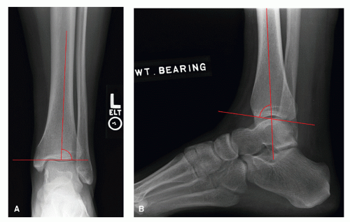
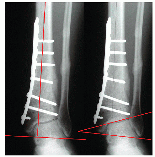
Radiographic imaging must include accurate weight-bearing orthogonal projections of the ankle and foot. The importance of positioning of the extremity during the radiographic process cannot be underestimated. Malpositioned or suboptimal technique will spuriously alter the apparent deformity and surgical plan accordingly. The anterior-posterior (AP) and lateral projections help determine the type and degree of deformity in all cardinal body planes. The center of the ankle joint relative to the long axis of the tibia is determined by subtending the middiaphyseal line from the distal tibia through the talus. In the normal ankle, the middiaphyseal line should pass through the center of the talus. On the lateral projection, the middiaphyseal line should pass through the superior talar dome and lateral process (Fig. 114.2). Additional radiographic parameters include the joint orientation angles called the lateral distal tibial angle (LDTA) (89 degrees ± 3 degrees) and the anterior distal tibial angle (ADTA) (80 degrees ± 2 degrees) (18) (Fig. 114.2). The talocrural angle (82 degrees ± 3.6 degrees) (18) is the angle formed by a line connecting the tip of the medial and lateral malleolus and middiaphyseal line. Deformity in the distal tibia will influence this angle and is therefore not reliable. The plafond malleolar (9 degrees ± 4 degrees) (18) angle is the angle formed by the tip of the malleoli and the tibial plafond. This angle is more reliable in the presence of distal tibial deformity (Fig. 114.3).
 Figure 114.2 The center of the ankle joint relative to the long axis of the tibia is determined by subtending the middiaphyseal line from the distal tibia, through the talus. A: In the normal ankle, the middiaphyseal line should pass through the center of the talus. B: On the lateral projection, the middiaphyseal line should pass through the lateral process of the talus. The LDTA (89 ± 3 degrees) and the ADTA (80 ± 2 degrees) are formed with the tangent to the tibial plafond (21). |
 Figure 114.3 The talocrural angle (82 ± 3.6 degrees) (21) is the angle formed by a line connecting the tip of the medial and lateral malleolus and middiaphyseal line. Deformity in the distal tibia will influence this angle and is therefore not reliable. The plafond malleolar (9 ± 4 degrees) (21) angle is the angle formed by the tip of the malleoli and the tibial plafond. This angle is more reliable in the presence of distal tibial deformity (21). |
The amount of translation of the calcaneus relative to the tibial axis and the angulation of the calcaneus in the frontal plane with respect to the tibia are two important criteria to evaluate. Since the translational deformity is the most overlooked, hindfoot alignment and long leg calcaneal axial
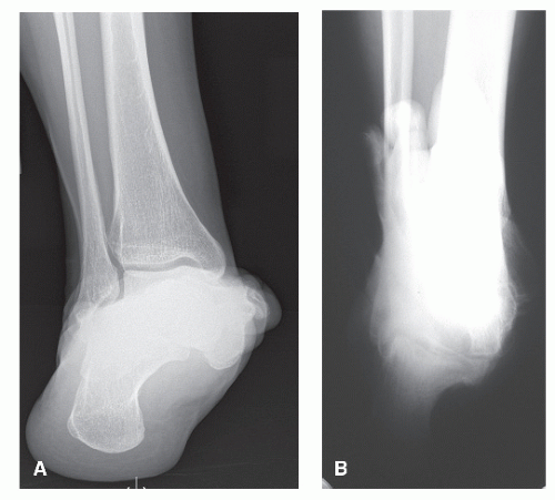
views can help determine these axial relationships of the foot to the lower leg. The hindfoot alignment view allows evaluation of the position of the calcaneus with respect to the ankle and distal tibia. The calcaneus is normally translated 1 cm lateral to the tibial middiaphyseal line (14). Long leg calcaneal axial views allow assessment of the subtalar joint and the relationship of the calcaneus to the anatomic axis of the tibia (Fig. 114.4). Infrequently, full-length standing films or stress views of the ankle are indicated. Advanced imaging such as MRI or CT may also be helpful to evaluate focal articular damage and periarticular pathology.
views can help determine these axial relationships of the foot to the lower leg. The hindfoot alignment view allows evaluation of the position of the calcaneus with respect to the ankle and distal tibia. The calcaneus is normally translated 1 cm lateral to the tibial middiaphyseal line (14). Long leg calcaneal axial views allow assessment of the subtalar joint and the relationship of the calcaneus to the anatomic axis of the tibia (Fig. 114.4). Infrequently, full-length standing films or stress views of the ankle are indicated. Advanced imaging such as MRI or CT may also be helpful to evaluate focal articular damage and periarticular pathology.
 Figure 114.4 A: The hindfoot alignment view allows evaluation of the position of the calcaneus with respect to the ankle and distal tibia. The calcaneus is normally translated 1 cm lateral to the tibial middiaphyseal line (14). B: Long leg calcaneal axial views allow assessment of the subtalar joint and the relationship of the calcaneus to the anatomic axis of the tibia.
Stay updated, free articles. Join our Telegram channel
Full access? Get Clinical Tree
 Get Clinical Tree app for offline access

|


