Ankle Equinus


Ankle Equinus


Michael S. Downey

Jaclyn M. Schwartz


Equinus deformity of the ankle has been classically described as a limitation of passive ankle joint dorsiflexion to less than a right angle of the foot on the leg. Lengthening of the Achilles tendon in the toe walker is one of the oldest known orthopaedic procedures. The reason is that the deformity is the most common malposition of the lower extremity in children with spastic types of cerebral palsy. Delpech (1) is reported to have performed the first subcutaneous section of the Achilles tendon in 1816. Stromeyer (2) then repeated this subcutaneous tenotomy in 1831 and later performed it on Dr. William Little, who had a talipes equinovarus deformity resulting from infantile paralysis. Little then became the major advocate of the tenotomy for the treatment of equinus in cerebral palsy because of his own successful heel cord lengthening (3). Since Little’s time, many surgical procedures to correct the equinus deformity have evolved.

However, until recently, these traditionally orthopaedic procedures were performed almost exclusively on patients with the spastic type of cerebral palsy. Hibbs (4), in 1914, described a procedure to lengthen the Achilles tendon as an adjunct to the correction of pes valgo planus deformity. This was probably the earliest description of surgical elongation of the tendon in a nonspastic attitude. Harris and Beath (5) also made mention of this in their article in 1948. Hall et al (6) in 1967 were the first distinctly to report a congenitally short Achilles tendon. They noted a group of 20 patients of whom 13 had a significant equinus deformity of the ankle but otherwise appeared normal. Root et al (7) reemphasized the frequency of the nonspastic ankle equinus abnormalities and laid the biomechanical framework to explain many of the foot deformities associated with this deformity. Sgarlato et al (8,9), in the 1970s, then summarized the previous findings when they described ankle equinus, its correction, and its effect on associated foot disorders.


ANATOMY

In treating equinus, many authors have attempted to break down the etiologic and pathologic components of the deformity. It has become obvious from their observations that an appreciation of the normal and pathologic anatomy is a prerequisite to addressing the proper evaluation of equinus, its biomechanical effects, and its surgical correction. Thus, the following is a brief description of the regular anatomic features of the component muscles of the superficial posterior crural group, the joints across which they function, and their relationship during the gait cycle.

The superficial group of the posterior crural muscles is composed of the gastrocnemius, soleus, and plantaris muscles. The gastrocnemius and soleus together form a tripartite muscular mass that shares the Achilles tendon and hence is often termed the triceps surae. The muscles of the superficial group are each innervated by the tibial nerve (segmental innervation: sacral levels one and two) and are vascularized by the posterior tibial artery; otherwise, they have unique anatomic features (10).


GASTROCNEMIUS MUSCLE

The gastrocnemius muscle is the most superficial muscle of the group and forms the belly of the calf (Fig. 43.1). Its ovoid heads take origin from facets above the femoral condyles by strong, flat aponeurotic bands marginally placed on each head. These bands descend about two-thirds of the way down the superficial (posterior) portion of the muscle. The muscular heads arise from these bands, and their fibers pass obliquely deep (anteriorly) to their tendon of insertion. The medial head is broader and thicker than the lateral head and also extends farther distalward (10). A bursa, which occasionally communicates with the knee joint, is deep (anterior) to the aponeurotic band of the medial head, whereas the concomitant band of the lateral head sometimes contains a fibrocartilaginous sesamoid bone (flabella). The fascia cruris separates the superficial (posterior) surface of the muscle from the small saphenous vein and the peroneal communicating and sural nerves (10) (Fig. 43.2).


SOLEUS MUSCLE

The soleus muscle lies deep (anterior) to the gastrocnemius and is broad and flat (Fig. 43.3). It arises from the back of the head of the fibula, the upper third of the posterior surface of the fibula, and the soleal line and the middle third of the tibia. Thus, like the gastrocnemius, the soleus has fibular and tibial heads that, although much flatter, arise from broad aponeuroses. These aponeuroses unite on the deep (anterior) surface of the muscle to form an arch over the posterior tibial vessels and the tibial nerve. The main portion of the muscle is formed by fibers arising from the superficial (posterior) surface of these aponeurotic bands. Descending in an oblique posterior course, the muscular fibers insert in bipenniform fashion into the deep (anterior) portion of the Achilles tendon (11). Occasionally, some muscle fibers arise from the deep (anterior) surface of the aponeuroses. These have been reported to have multiple routes of insertion and rarely can insert separately from the Achilles tendon (12,13,14,15,16 and 17). The deep transverse fascia separates the soleus from the underlying flexor hallucis longus, flexor digitorum longus, tibialis posterior, and the aforementioned posterior tibial vessels and tibial nerve (10).


Figure 43.1 Muscles of the right calf: posterior view of the superficial layer.


PLANTARIS MUSCLE

Although much smaller than the gastrocnemius and soleus muscles, the plantaris muscle must also be considered in a discussion of the superficial posterior crural muscles. The plantaris arises in close association with the lateral head of the gastrocnemius. It has a small, flat fusiform belly that, after 7 to 10 cm, ends as a long, slender tendon. The tendon passes obliquely between the gastrocnemius and soleus and runs along the medial edge of the Achilles tendon to be inserted with it. The plantaris, more so than the other two muscles, is extremely variable in origin, structure, and insertion (10). It has been reported to be absent in about 7.05% of all cases (18) (Fig. 43.4). Although only a rudimentary muscle, the plantaris is generally considered to function much like the gastrocnemius and, as such, must be addressed during many surgical procedures for equinus.


Figure 43.2 Cross section through the lower third of the right leg.


Figure 43.3 Muscles of the right calf: posterior view of the superficial layer with the gastrocnemius excised.


ACHILLES TENDON

The Achilles tendon, the thickest and strongest human tendon, serves as the insertion for both the gastrocnemius and soleus and occasionally the plantaris. It is approximately 15 cm in
length and begins near the middle of the leg. The tendon becomes most rounded at a level roughly 4 cm proximal to its insertion and then expands again to attach to the posterior middle third of the calcaneus (10). Sarrafian (19) described a more expansive insertion, including the entire posterior distal half of the calcaneus, as the tendon’s normal insertions. Although complete anatomic separation of the gastrocnemius and soleus components of the tendon is not possible, comments about the course of the majority of their fibers can be made.


Figure 43.4 Variations of tendinous insertion of the plantaris muscle.

The gastrocnemius portion of the Achilles tendon begins as a broad aponeurosis from the deep (anterior) surface of the muscle bellies. Its fibers converge and rotate at the same time toward the lateral side of the soleal component. At the insertion of the Achilles tendon, the gastrocnemius component usually comprises the lateral side of the superficial (posterior) surface and a small portion of the lateral aspect of the deep (anterior) surface of the tendon.

The soleus portion, meanwhile, is thicker than that derived from the gastrocnemius. It begins as an aponeurotic band from the superficial (posterior) portion of the muscle and continues to receive tendinous components as it proceeds inferiorly. Opposing the gastrocnemius, it rotates toward the medial side and at the tendon’s insertion generally comprises the medial two-thirds of the deep (anterior) surface and a small portion of the medial side of the superficial (posterior) surface (11) (Fig. 43.5). White was one of the first to describe this twisting of the tendon and advocated its knowledge for his surgical procedure (20). Morimoto and Ogata (21) hypothesized that the torsion was nature’s way of increasing the tendon’s architectural strength during weight-bearing. Conversely, a few authors, including Mercado (22), suggested that the fibers of the Achilles tendon do not rotate at all but merely appear oblique. Most likely, the tendinous contributions of the gastrocnemius and soleus are not entirely separable, and the number and strength of the fibers exchanged and the degree of torque in the tendon vary greatly (11,23).


BIOMECHANICS

When one considers the origin of an ankle equinus deformity, the joints across which these muscles function are also of prime importance. The gastrocnemius and plantaris muscles originate from the femur and cross the knee joint, whereas the soleus muscle, with its tibial and fibular origins, does not. The tendinous components of all three muscles then cross both the ankle and subtalar joints. Therefore, the gastrocnemius and plantaris can be accurately termed three-joint muscles and the soleus a twojoint muscle. This differentiation assumes clinical importance and is the basis of the Silfverskiold test (24), as discussed later.


Figure 43.5 Achilles tendon, with the most common variations of gastrocnemius and soleus components at the level of insertion into the calcaneus. The gastrocnemius contribution is unshaded; the soleus contribution is shown in green.


Figure 43.6 Illustrations demonstrating the changing orientation of the subtalar joint axis when the joint is supinated (A), neutral (B), or pronated (C).

Occasionally, the gastrocnemius and plantaris are inaccurately described as two-joint muscles and the soleus as a onejoint muscle. This misconception arose when many authors considered the muscular action of the group across the subtalar joint as insignificant. Indeed, the neutral subtalar joint axis is comparatively parallel to the Achilles tendon, whereas the axes of the ankle and knee are relatively perpendicular to the tendon (7). However, when the subtalar joint pronates, its axis becomes more perpendicular to the Achilles tendon and therefore must be considered in a discussion of equinus (see later discussion of compensated equinus) (Fig. 43.6).

Although the joints across which the muscles of the superficial posterior crural group function must be remembered, the relation and attitude of the muscles to these joints during the gait cycle are of utmost importance in understanding the deformity. Root et al (7) stated that the gastrocnemius and soleus muscles normally begin to contract toward the end of the contact period of the stance phase of gait (Fig. 43.7). They continue to contract throughout the midstance period and through the first portion of the propulsive period. They are normally inactive throughout the remainder of the gait cycle (Fig. 43.8). Thus, by virtue of their origin, the gastrocnemius is involved in knee flexion, and both muscles are then involved in heel lift. Just before heel lift, with the knee still fully extended and the subtalar joint in its neutral position, the maximum amount of ankle joint dorsiflexion is required (Fig. 43.9).


ETIOLOGY AND CLASSIFICATION

When an ankle joint equinus has been identified, its etiologic and associated compensatory pathologic features must then be determined. In the past, many authors offered their definitions and classifications of equinus along with their recommended corrective procedures. Until recently, however, few had attempted to break down the equinus deformity into specific forms and etiologic entities.

McGlamry and Kitting (29), in 1973, classified and provided clinical definitions of five etiologic types of equinus foot: talipes equinus, osseous equinus, gastrocnemius-soleus equinus, metatarsal equinus, and forefoot equinus. Their definition encompassed more than ankle joint equinus because they described the equinus foot as “one which at rest drops below the dangling foot to leg angle. This excessive plantarflexion of the foot may occur at the ankle, but often occurs at the midtarsal joint or at the tarsometatarsal joint.” Obviously, this definition includes types of equinus occurring distal to the ankle joint. Metatarsal equinus is a state of plantarflexion of the forefoot in relation to the rearfoot at the tarsometatarsal or Lisfranc joint. Forefoot equinus is a plantarflexed attitude of the forefoot on the rearfoot at the midtarsal or Chopart joint. Both are examples of anterior cavus or equinus occurring in the foot and not in the ankle.

Ankle equinus occurs at the ankle joint and may result from muscular deformity, osseous deformity, or a combined muscular-osseous deformity. Thus, ankle equinus is most easily classified into these forms with further subdivision into etiologic types (30) (Table 43.1).


MUSCULAR FORMS

The posterior crural muscles individually, in part or as a whole, may be in a state of contracture and may thus produce an ankle equinus deformity. These contractures may be spastic or nonspastic (i.e., congenital, idiopathic, or acquired shortness).

Spastic equinus is the oldest known form of ankle joint equinus. It is usually found in patients with neuromuscular diseases such as cerebral palsy. The posterior crural musculature may be partially or wholly spastic and can overpower the weaker anterior musculature entirely. Hypertonicity of the involved musculature, hyperreflexia, and clonus are often found to be present. In this manner, pyramidal or corticospinal level spasticity may be differentiated from other motor disorders (Table 43.2). In addition, efforts should be made to determine the muscle

or muscles that are spastic inasmuch as this evaluation aids in proper treatment. As previously mentioned, in normal gait, the gastrocnemius and soleus muscles primarily function during the stance phase of the gait cycle. The anterior musculature, or dorsiflexors, are active mainly during the swing phase of the gait cycle. This, however, is not true in the presence of spastic equinus because the involved posterior crural musculature is spastic throughout the entire gait cycle. Depending on the degree of spasticity, the posterior group may overpower the anterior group. The gait is then no longer heel-toe but becomes toe-toe. This is the classic toe walker, as described in the earliest literature. Lesser degrees of spasticity may allow the heel to come down or even contact the floor (toe-heel gait) (31). Generally, the spastic forms of ankle equinus are more resistant to corrective treatment than are nonspastic forms.


TABLE 43.1 Types of Ankle Joint Equinus


Muscular Forms


Osseous Forms


Gastrocnemius equinusa


Talotibial exostosesa


Spastic


Anterior


Nonspastic


Posterior


Other


Gastrocnemius-soleus equinusa


Tibiofibular syndesmosis limitationa


Pseudoequinusa


Spastic


Combination formsa


Nonspastic


a One of six major etiologic types.


TABLE 43.2 Motor Disorders


Spinomuscular


Muscular


Neural


Spinal


Extrapyramidal


Pyramidal


Cerebellar


Psychomotor


Loss of motor power


Focal-segmental


Focal-segmental


Focal-segmental


Generalized


Generalized


None


No true loss


Usually proximal and axial muscle groups


Usually distal limb musculature


Usually distal limb musculature


Entire limb and movements


Entire limb and movements


Ataxia may simulate loss of power


Bizarre, may simulate any type


Tone


Complete Flaccid


Complete Flaccid


Complete Flaccid


Incomplete Rigid


Incomplete Spastic


Hypotonic (ataxia)


Normal or variable, may be increased


Atrophy


Present


Present


Present


Absent


Minimal (due to disuse and chronic paresis)


Absent


Absent


Fasciculations


May be present


Absent


May be present


Absent


Absent


Absent


Absent


Reaction of degeneration


Present


Present


Present


Absent


Absent


Absent


Absent


Electromyogram


Interference pattern


Normal until late in disease


Reduced


Reduced


Fibrillation potential


Not usually present


Present


Usually present


Action potential


Short duration


Prolonged with normal or polyphasic potentials


Prolonged with occasional giant potentials


Evoked sensory and mixed nerve potentials


Normal


Absent, diminished amplitude, or prolonged conduction time


Normal


Reflexes


Deep


Diminished and preserved until late


Absent early


Absent early


Normal or variable


Hyperactive


Diminished or pendular


Normal or increased range


Superficial


Diminished


Absent


Absent


Normal or increased


Diminished or absent


Normal


Normal or increased


Pyramidal tract response


No


No


No


No


Yes


No


No


Sensory deficit


Absent


Usually present


Absent


Absent


May be present (stereognosis or other cortical)


Absent


Absent


Trophic disturbance


Present


Present


Present


Absent


Usually absent


Absent


Absent


Ataxia


Absent


Absent


Absent


Absent


Absent


Present


Absent (may simulate ataxia)


Abnormal movements


Absent


Absent


Absent


Present


None


May be present (intention tremor and ataxia)


May be present


Associated movements


Normal


Normal


Normal


Absence of normal associated movements


Presence of pathologic associated movements


Normal


Normal


Modified from DeJong RN. The neurological examination, 3rd ed. New York: Harper & Row, 1967:382.


Today, nonspastic muscular ankle equinus is discussed with increasing frequency and is now considered to be more common than spastic muscular ankle equinus. Congenital nonspastic shortness of the posterior crural musculature that creates an ankle equinus deformity was first reported by Hall et al (6) in 1967. Toe walking may be considered normal when a child first begins to walk. However, within a 3- to 6-month period, or by age 7 years, the child should discontinue a toe-toe or toe-heel gait pattern and should learn to walk with a heel-toe gait. Children with a congenital shortness of the gastrocnemius or gastrocnemius-soleus complex may exhibit toe-toe gait but are able to lower their heels to the ground. The toe walking may be considered habitual when the child is capable of heel-toe gait, but prefers toe-toe or toe-heel gait. Occasionally, with examination or on surgical correction, an abnormally low insertion of the soleus muscle or an accessory soleus muscle may be identified (32). Generally, however, no anomalies are identified, and results of the neurologic examination are completely normal. Thus, the diagnosis of congenital or idiopathic nonspastic ankle equinus is one of exclusion, in which the only abnormality is a limitation of ankle joint dorsiflexion (33,34,35 and 36). In addition, several authors have described hereditary nonspastic gastrocnemius-soleus contractures as an autosomal dominant condition with variable severity of expression (37,38). More recently, Sobel et al (39) analyzed 60 idiopathic toe walkers, and concluded that treatment should be initiated because fixed contractures may result with time.

In addition to congenital nonspastic shortness, acquired shortness of the posterior crural musculature creating an ankle equinus can result from nonspastic causes. Davis law states that “soft tissue under prolonged tension will elongate” and that the “same is true in retrospect.” Soft tissue contracts to its altered position, and this is the underlying etiologic factor in most cases of acquired muscular ankle equinus. Situations that create prolonged or repeated plantarflexed or equinus positions of the ankle may result in ankle equinus caused by muscular contracture. Common examples range from prolonged casting with the ankle joint plantarflexed to the repetitive use of highheeled shoes. Acquired shortness may also result from trauma (e.g., an Achilles tendon rupture with healing in a shortened position) or iatrogenic causes.

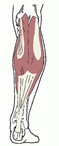
Although all or any portion of the posterior crural musculature may contribute to a muscular form of ankle equinus, gastrocnemius and gastrocnemius-soleus contractures are considered to be the most common. If a muscular ankle equinus deformity is suspected, the clinician must first determine whether the deformity is spastic or nonspastic by performing a complete neurologic history and physical examination. In patients with spasticity, studies have discussed the use of a tibial nerve block to help to differentiate the amount of fixed contracture from spastic contracture (40,41). These investigators measured the amount of torque about the ankle joint before and after the tibial nerve block. They theorized that the contracture present before the block was a result of both the spasticity and fixed contracture, and the contracture present after the block was the fixed contracture alone. This hypothesis may have future implications for the choice of surgical procedures or treatment to be performed.

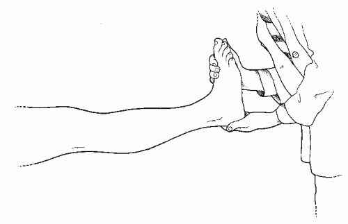
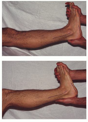
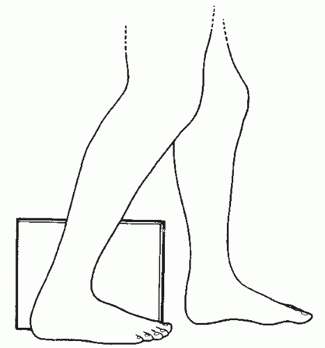
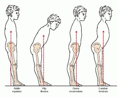
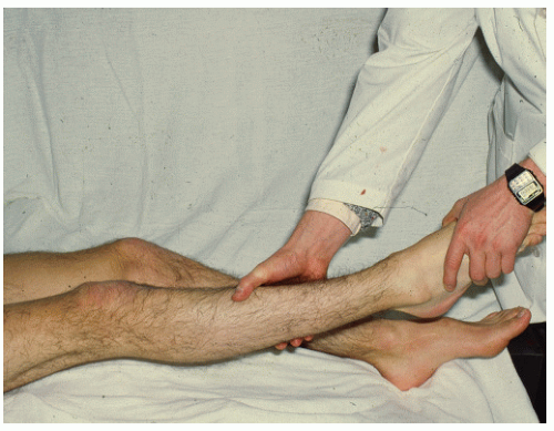
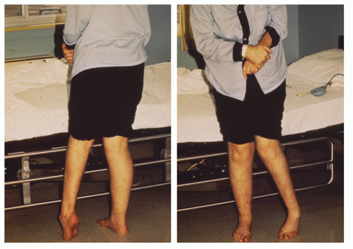
Once an assessment for the presence or absence of spasticity is completed, the examiner may evaluate the patient for the specific muscles involved. This later examination begins by confirming the presence of ankle or posterior equinus. With the patient lying flat and in a supine position, the leg is elevated slightly from the supporting surface with the knee maintained in an extended position (Fig. 43.11). If genu recurvatum is found to be present, the measurement is made in that position, because that is the attitude in which the knee functions just before heel lift. The examiner then grasps the patient’s foot and maintains the subtalar joint in a neutral position (or a slightly supinated position, not pronated). Appropriate dorsiflexory pressure is then applied to the foot at the ankle joint, and the angle formed by the bisection of leg to the rearfoot in the sagittal plane is measured. If the amount of ankle joint dorsiflexion is less than 10 degrees (or greater than an 80-degree angle of foot to leg), a posterior or ankle equinus has been identified.


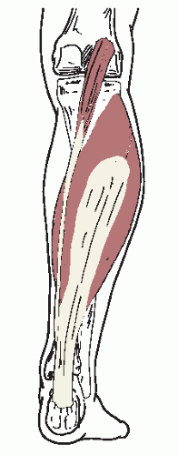
Gastrocnemius Equinus

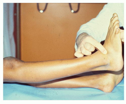
Gastrocnemius equinus is an etiologic type of ankle equinus caused by contracture or shortening of the gastrocnemius or plantaris muscles. Once a posterior or ankle equinus has been identified, the 
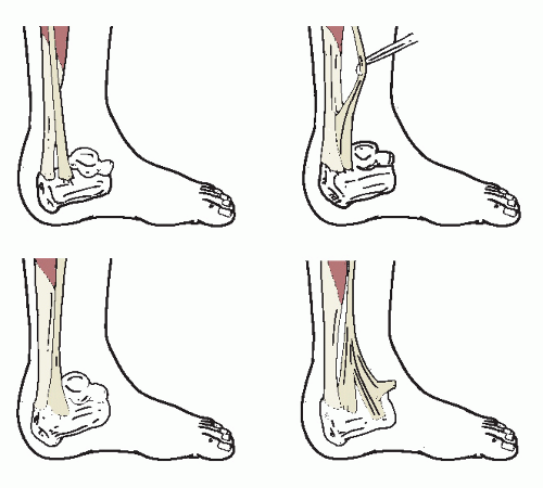
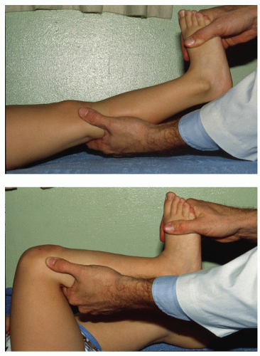
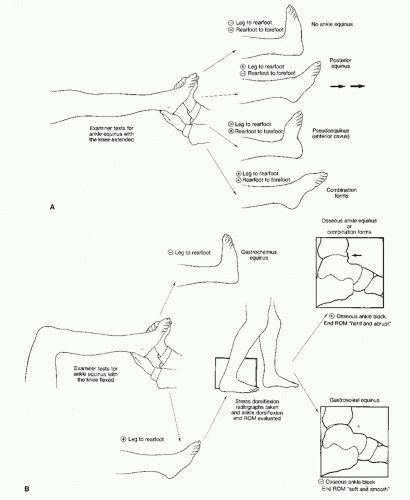
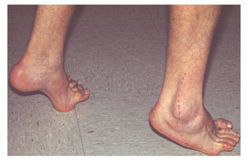
Silfverskiold test may then be performed to differentiate gastrocnemius equinus from the remaining types of posterior equinus (24). With the patient remaining flat and in the supine position, the examiner flexes the patient’s knee to a right angle and again dorsiflexes the patient’s ankle while making certain to maintain the subtalar joint in a neutral or slightly
supinated position (
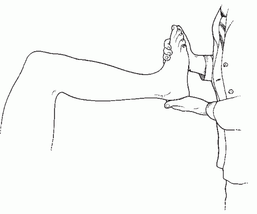
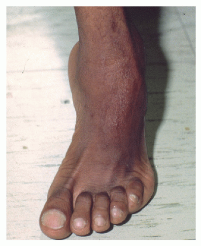
Fig. 43.12). As before, a measurement of the sagittal plane angular relation of the bisection of the leg to the rearfoot is taken. If more than 10 degrees of ankle joint dorsiflexion (or less than an 80-degree angle of foot to leg) is found, a gastrocnemius equinus has been diagnosed. The gastrocnemius and plantaris muscles cross the knee joint, unlike the soleus muscle. When the knee is flexed, the courses of the gastrocnemius and plantaris are shortened, and the muscles are relaxed when the ankle joint is dorsiflexed. Conclusively, then, if there is a limitation of ankle joint dorsiflexion with the knee extended, but not with the knee flexed, the limitation can be attributed to tight gastrocnemius and plantaris muscles (Fig. 43.13). If, after the Silfverskiold test is performed, a limitation of ankle joint dorsiflexion is still found to be present, other sources of ankle joint equinus must be present.


Figure 43.11 Initial clinical examination for ankle joint equinus.


Figure 43.12 Silfverskiold test: clinical examination for gastrocnemius equinus.


Gastrocnemius-Soleus (Gastrocsoleus) Equinus

Gastrocnemius-soleus (gastrocsoleus or gastrocsoleal) equinus is an etiologic type of ankle joint equinus caused by contracture or shortening of all or some of the soft tissue structures passing posterior to the ankle joint axis, including the components of the triceps surae. These sources may include any soft tissue structures that cross the ankle joint and not the knee joint. These structures, of course, must be capable of limiting ankle joint dorsiflexion and include the soleus muscle, the peroneal muscles (longus and brevis), the flexor longus muscles (digitorum and hallucis), the tibialis posterior muscle, and the ligamentous and capsular tissue of the ankle joint. Tightness and restriction by any of these structures may cause a limitation of ankle joint dorsiflexion with the knee in both the flexed and extended positions. Further, these conditions may cause a concurrent gastrocnemius (and plantaris) tightness. The tightness cannot be clinically isolated because of the limitation of ankle joint dorsiflexion with the knee flexed and extended and the
inconclusiveness of the Silfverskiold test. Consequently, most examiners assume that the gastrocnemius and plantaris muscles are tight when these other soft tissue structures are restrictive (42). Gastrocsoleus equinus is the most common example of posterior equinus demonstrating a limitation of ankle joint dorsiflexion with the knee both flexed and extended. It must, however, be differentiated from osseous ankle equinus, which produces similar findings (Fig. 43.14).


Figure 43.13 Clinical examination demonstrating gastrocnemius equinus. Note the limitation of ankle joint dorsiflexion with the knee extended, but adequate dorsiflexion with the knee flexed.


Figure 43.14 Clinical examination demonstrating gastrocsoleus equinus or osseous equinus. Note the limitation of ankle joint dorsiflexion with the knee extended or flexed.


OSSEOUS FORMS

Osseous pathologic changes about the ankle may also limit ankle joint dorsiflexion and may cause an osseous form of ankle equinus. In addition, osseous equinus within the foot can cause the false appearance of an ankle joint equinus. This pseudoequinus is discussed and is differentiated as a form of osseous equinus.


Talotibial Exostoses

Talotibial exostoses represent an etiologic type of ankle equinus and include any osseous projections from the margins of the trochlear surface of the talus and the distal articular surface of the tibia that could limit ankle joint dorsiflexion. Most commonly, these projections manifest as anterior talotibial exostoses that directly limit ankle joint dorsiflexion. However, posterior talotibial exostoses or, more rarely, other pathologic conditions about the ankle joint (e.g., osseous bridging between the tibia and fibula in the distal syndesmotic area) can also limit ankle joint dorsiflexion. This etiologic type of equinus demonstrates clinically limited ankle joint dorsiflexion with the knee both extended and flexed.

Although the range of ankle joint dorsiflexion is limited with both talotibial exostoses and gastrocsoleus equinus, the end range of dorsiflexory motion is soft or smooth in gastrocsoleus equinus and is hard or abrupt and firm in osseous ankle equinus caused by talotibial impingement. This observation is highly subjective, and considerable clinical experience is required to establish the correct diagnosis consistently without further evaluation.

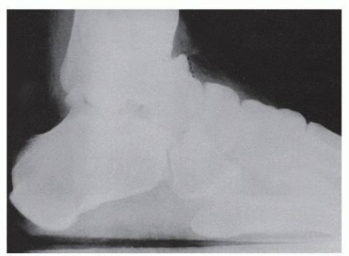
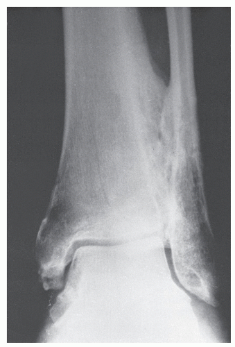
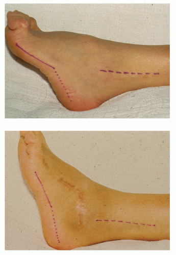
Generally, additional evaluation is desired, and radiographs should be taken to eliminate the possibility that a bony blockage is associated with a talotibial exostosis. A conventional lateral radiograph of the ankle may be taken and examined for possible osseous projections from the margins of the trochlear surface of the talus or distal surface of the tibia that could limit the range of ankle joint dorsiflexion (Fig. 43.15). In some instances, a tibial exostosis is seen along with a depression in the dorsal talar neck to accommodate the anterior tibial prominence. This has been described as the divot sign, because the divot or depression occurs in the dorsal talar neck instead of a corresponding talar exostosis (43). Additionally, synostoses may rarely occur between the tibia and fibula limiting ankle joint dorsiflexion. If this condition is suspected, a conventional mortise or medial oblique view of the ankle can be used to evaluate the distal tibiofibular articulation for any osseous pathologic changes (Fig. 43.16). Further, a lateral weight-bearing radiograph of the ankle may be taken, with the patient instructed to bend the knee forward with as much force as possible (creating ankle joint dorsiflexion) while keeping the heel on the ground (Fig. 43.17). This view, also referred to as a charger view or stress-dorsiflexion view, of the ankle may also be taken with the patient in a non-weight-bearing attitude. To obtain the view in a non-weight-bearing position, the examiner simply performs the aforementioned Silfverskiold test, and a lateral radiograph of the ankle is taken. Quantitatively, the sagittal plane angulation of the long axis of the tibia to the long axis of the talus is
measured in both the standard lateral and stress-dorsiflexion radiographs. If a limitation of ankle joint dorsiflexion is present in either view and a bony projection of tibia, talus, or other osseous structure is found to limit further dorsiflexion of the ankle joint, then a diagnosis of osseous ankle equinus can be made. If no apparent bony projections are present, a muscular gastrocsoleus ankle equinus with a tight triceps surae complex or associated posterior structures must be assumed (8).


Figure 43.15 Radiograph demonstrating anterior talotibial exostoses causing osseous ankle equinus.


Figure 43.16 Osseous bridging of the distal tibiofibular articulation. Restriction of normal function and movement of syndesmosis can cause ankle equinus.


Figure 43.17 Stress dorsiflexion or charger view of the ankle joint.


Pseudoequinus

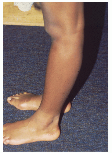
Anterior equinus, also known as anterior cavus, is a deformity represented by a plantarflexed attitude of the forefoot or any of its component parts. This type of equinus may be subdivided into four types based on the apex of the deformity in the sagittal plane. These types include the aforementioned metatarsal equinus and forefoot equinus along with lesser tarsal equinus (an excessive plantarflexed deformity occurring in the lesser tarsal bones themselves) or a combination of these deformities (42). Although these types of anterior equinus do occur in the foot and not in the ankle, an apparent or false ankle equinus mistakenly can be measured clinically. To explain, ankle equinus is a sagittal plane measurement of the bisection of the leg to the rearfoot. The inexperienced clinician often falsely identifies an ankle equinus in the anterior cavus foot if he or she measures the sagittal plane bisection of the leg to the forefoot. This allows any plantarflexion of the forefoot on the rearfoot to be included in the measurement.

Identifying this problem and to avoid confusion, Whitney and Green (42) appropriately coined the term pseudoequinus to account for the false ankle equinus seen in an anterior cavus foot type as previously described. Thus, pseudoequinus is an angular relation of equinus of the forefoot to the leg in the sagittal plane without an equinus relation of the rearfoot to the leg in the sagittal plane. It is most commonly associated with a structural anterior equinus (i.e., anterior cavus) deformity. Pseudoequinus is not a true ankle joint limitation but an excessive demand for ankle dorsiflexion created by forefoot equinus. This occurs because the entire foot must be dorsiflexed at the ankle to allow the heel of the foot with anterior equinus to reach the ground. Thus, ankle joint dorsiflexion is overtaxed to compensate for the anterior equinus deformity (42). Surgical correction of pseudoequinus is primarily by osseous correction to decrease the angular equinus relationship of the forefoot to the rearfoot (Fig. 43.18). These surgical procedures are discussed elsewhere in this text.


COMBINATION FORMS

Perhaps one of the least understood forms of ankle equinus is that occurring as a combination of the aforementioned types. Unless the clinician is particularly attentive, these forms are often overlooked.

Pseudoequinus may occur in combination with any of the other forms of ankle equinus. If the examiner notes a sagittal plane equinus relation of both the rearfoot to the leg and the forefoot to the rearfoot, then such a relationship exists. Although this combined relationship is rare, it does occur, and one should not presume that the cavus foot cannot have an associated muscular or osseous ankle equinus. Once the cause of the anterior equinus is identified, the examiner may perform the tests used to differentiate the isolated ankle equinus deformities to determine whether the pseudoequinus is occurring in
combination with a muscular form of equinus (i.e., gastrocnemius or gastrocsoleus equinus) or a talotibial exostosis (44).


Figure 43.18 Demonstration of pseudoequinus before and after triple arthrodesis with digital stabilizations. Note the marked decrease of sagittal plane forefoot to rearfoot equinus relationship after surgical correction.

Talotibial exostoses may also occur in combination with the other types of equinus. They can occur with a pseudoequinus. To determine an osseous equinus in combination with a gastrocnemius or gastrocsoleus equinus, one must first surgically resect the osseous block. Once the osseous block has been removed, the surgeon may then reevaluate the patient for muscular ankle equinus. If one finds 10 degrees of ankle joint dorsiflexion to be present with the knee fully extended, the osseous equinus may be assumed to be an isolated deformity. Often, however, the posterior musculature has adapted, and a gastrocnemius or gastrocsoleus equinus is discovered. Obviously, the Silfverskiold test can then be used to differentiate these muscular forms of ankle equinus (44).

In summary, a stepwise approach can be used in the etiologic evaluation of ankle equinus (Fig. 43.19). The clinician must be aware that equinus of the ankle often has more than one cause.


SUBJECTIVE AND OBJECTIVE ASSOCIATED PATHOLOGIC FINDINGS

Pathologic changes within the lower extremity can occur if adequate dorsiflexion is not available at the ankle joint. In many instances, these pathologic changes appear as both subjective patient complaints and objective compensatory findings. These subjective and objective changes may be divided into those occurring proximal and distal to the ankle joint.


PROXIMAL ASSOCIATED PATHOLOGIC CHANGES

With an ankle equinus deformity, the patient’s center of gravity is shifted slightly posterior as compared with normal. Proximal pathologic changes occur in an attempt to reposition the center of gravity anteriorly to a more normal position. Representative proximal compensatory changes include lumbar lordosis, hip flexion, knee flexion, and genu recurvatum (Fig. 43.20). Hibbs (4) accurately noted the symptoms associated with these deformities, stating that “these patients suffer from excessive fatigue, pains in the legs often referred to the back, nervousness, and mental lassitude”. Indeed, lower back strain or pain, hip pain, knee pain, and calf cramping and fatigue are postural symptoms commonly associated with these proximal changes.

Lumbar lordosis or saddle back frequently occurs with severe ankle equinus and is usually associated with hip and knee flexion. These three deformities combine to enable the patient to assume a crouched position and to shift the center of gravity forward. Although these deformities often occur together, they may also occur in pairs or alone. In addition, hamstring tightness is common with these deformities, and the clinician evaluating an ankle joint equinus must also assess the hip and thigh musculature. The biceps femoris, semitendinosus, and semimembranosus, often grouped familiarly as the hamstring group, span the hip and knee joints, integrating extension at the former and flexion at the latter. Thus, the patient with tightness or spasm of the hamstrings may exhibit a flexion deformity at the knee. The gastrocnemius muscle, as previously mentioned, also crosses the knee joint. If a patient with an ankle joint equinus has a knee flexion deformity, the clinician must attempt to differentiate whether the cause involves the hamstring or gastrocnemius muscle (45).

To perform this evaluation, the clinician attempts to extend the patient’s knee fully with the ankle joint maximally plantarflexed (Fig. 43.21). This places the gastrocnemius muscle in a relatively relaxed position while having no direct effect on the hamstring group. If the knee joint cannot be fully extended, the flexion deformity is generally not attributable to the gastrocnemius. Conversely, if the knee can be fully extended, then the flexion deformity is attributed to the gastrocnemius. When the gastrocnemius is extremely tight or spastic, the muscle may still be tight with the ankle maximally plantarflexed. The clinician can usually identify this circumstance by simply palpating for tightness of the Achilles tendon while performing the test. The tendon should not be tight with the ankle joint maximally plantarflexed if the gastrocnemius is not a contributing force to the knee flexion deformity.

Genu recurvatum, or a position of knee joint hyperextension, is another proximal compensatory change often seen with an ankle equinus deformity (Fig. 43.22). The knee can compensate for a posteriorly displaced center of gravity by hyperextension that brings the center of gravity forward (32). Genu recurvatum is a functional deformity, and an initial clinical examination for ankle joint dorsiflexion must have the patient positioned with the knee fully extended.


Figure 43.19 Stepwise approach to ankle joint equinus.


Figure 43.20 Proximal compensatory mechanisms of ankle equinus.


DISTAL ASSOCIATED PATHOLOGIC CHANGES

The foot can compensate for an ankle joint equinus only after pronatory events allowing the forefoot to dorsiflex on the rearfoot have occurred. Specifically, subtalar joint pronation followed by midtarsal joint pronation must occur. Both the subtalar joint and the midtarsal joint have axes about which the motion is supinatory (inversion, adduction, plantarflexion) and pronatory (eversion, abduction, dorsiflexion). Further, the midtarsal joint possesses two axes—talonavicular and calcaneocuboid—and has both an oblique axis and a longitudinal axis (Fig. 43.23). Because of the orientation of the axes, the dominant motions about the subtalar joint axis and longitudinal axis of the midtarsal joint are inversion and eversion. In contrast, dorsiflexion and plantarflexion are the prevalent planes of motion about the oblique axis of the midtarsal joint. The extent of congruous surface areas between the talonavicular and calcaneocuboid joints determines the range of motion about the oblique axis of the midtarsal joint. When the joint surfaces are incongruous (axes become oblique), the midtarsal joint is said
to be locked, and motion in both a supinatory and pronatory direction ceases. When the joint surfaces are congruous (axes become parallel), the midtarsal joint becomes unlocked, and supinatory-pronatory motion can occur (7).


Figure 43.21 Clinical evaluation to determine the cause of a knee flexion deformity.


Figure 43.22 Genu recurvatum (hyperextension of knee) to compensate for ankle equinus deformity.


Figure 43.23 Midtarsal joint axes of motion: oblique and longitudinal. (Redrawn after Root ML, Orien WP, Weed JH, eds. Normal and abnormal function of the foot: clinical biomechanics, vol. 2. Los Angeles, CA: Clinical Biomechanics, 1977:42.)

Elftman (46) was the first to explain the effect of subtalar joint position on the range of motion at the midtarsal joint. He described a congruity (or parallelism) that develops between the axes of the talonavicular and calcaneocuboid joints when the subtalar joint is pronated and an incongruity (or obliquity) that develops when the subtalar joint is supinated. Thus, when the subtalar joint pronates, the midtarsal joint unlocks and pronates itself. This translates to dorsiflexion about the oblique axis of the midtarsal joint because this is the dominant motion associated with pronation about that axis (Fig. 43.24). This dorsiflexion of the forefoot on the rearfoot compensates for limited ankle joint dorsiflexion. Sgarlato (8) divided this distal compensation into three groups, each with specific associated pathologic findings: (a) uncompensated or absence of distal compensation, (b) fully compensated or full distal compensation, and (c) partially compensated.


Figure 43.24 Illustrations demonstrating the change of orientation of the oblique midtarsal joint axis with pronation and supination of subtalar joint. The oblique midtarsal joint axis is capable of more dorsiflexion and plantarflexion with subtalar joint pronation.


No Distal Compensation

Ankle equinus deformity may occur with minimal distal compensation in which the subtalar joint remains supinated, and minimal or no subtalar and midtarsal joint motion occurs. Therefore, a lack of dorsiflexion at the ankle joint cannot be compensated for by abnormal subtalar and midtarsal joint pronation. Sgarlato (8) stated that fewer than 1% of all patients with ankle equinus belong in this category. Such patients usually have severe neuromuscular disease (e.g., cerebral palsy), spastic musculature, and an associated severe equinovarus and anterior equinus type foot (Fig. 43.25). Proximal compensation
typically occurs because the distal compensation is minimal or absent.


Figure 43.25 Patient with spastic hemiplegia.


Figure 43.26 Toe walking secondary to spastic diplegia.

Commonly, this patient is the true toe walker with no heel contact, and the forefoot, or ball of the foot, bears the weight during stance and gait (Fig. 43.26). Obviously, this type of gait is unstable. Submetatarsal head tylomas may certainly be expected because of the increased declination of the metatarsals and their prominence during weight-bearing. Further, digital contractures can be seen because of the extensor substitution phenomenon associated with this type of drop foot and the need for additional dorsiflexion at the ankle joint (Fig. 43.27). Occasionally, symptoms of plantar fasciitis or posterior knee pull syndrome are present (42). Other deformities such as hallux abducto valgus, commonly seen with hypermobile pronatory foot types, are generally absent (8).


Full Distal Compensation

In contrast, an ankle equinus deformity may occur with full distal compensation with subtalar joint and midtarsal joint pronation. A sufficient amount of dorsiflexion occurs at the oblique axis of the midtarsal joint to compensate fully for the limitation of ankle joint dorsiflexion. This is the hypermobile flatfoot with the rearfoot maximally everted to the floor and the forefoot everted on the rearfoot (Fig. 43.28). Many authors have advocated adjunctive Achilles tendon lengthenings in their corrective procedures for collapsing pes valgo planus deformity. The ankle equinus deformity with full distal compensation generally occurs with the most severe symptom complex and associated pathologic changes (8).


Figure 43.27 Severe anterior cavus foot type with associated digital contractures.

Only gold members can continue reading. Log In or Register to continue

Stay updated, free articles. Join our Telegram channel

Jul 26, 2016 | Posted by in MUSCULOSKELETAL MEDICINE | Comments Off on Ankle Equinus

Full access? Get Clinical Tree

Get Clinical Tree app for offline access