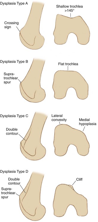
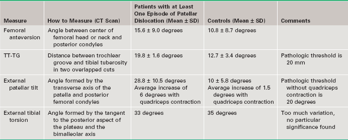
Chapter 89 Trochlear dysplasia is one of the major factors causing patellar dislocations (together with patella alta, excessive tibial tubercle–trochlear groove distance, and excessive patellar tilt). This anatomic abnormality is present in 96% of patients with patellar dislocations.1 Trochlear osteotomy or trochleoplasty is the procedure designed to correct the abnormal shape of the trochlea, improving patellar tracking and preventing instability. It is a logical procedure from the biomechanical point of view. Despite being technically demanding, it yields good results, and therefore it should be known to surgeons dealing with the patellar dislocation population. Trochleoplasty should be combined with other procedures to treat the other major causes of instability and is rarely performed alone. Imaging is essential to understand trochlear dysplasia and to allow its classification. Whereas normal trochleae have sulci of adequate depth, dysplastic trochleae are shallow, flat, or even convex. On lateral x-ray projections, this is represented by the crossing sign—the groove line reaches (or crosses) the line representing the superior edge of the facets. Two other features are typical of dysplastic trochleae on lateral views: the supratrochlear spur and the double contour sign. The supratrochlear spur is the same sometimes visualized in the surgical exposure, located in the superolateral aspect of the trochlea. It corresponds to an attempt to contain the lateral displacement of the patella. The double contour represents the medial hypoplastic facet, seen posterior to the lateral one in the lateral view. Based on these signs, and sometimes aided by computed tomography (CT) axial views, trochlear dysplasia may be classified into four types2: • Type A: Presence of crossing sign in the lateral true view. The trochlea is shallower than normal ones, but still symmetric and concave. • Type B: Crossing sign and trochlear spur. The trochlea is flat or convex in axial images. • Type C: Presence of crossing sign, and in addition the double-contour sign can be found on the lateral view, representing the medial hypoplastic facet. There is no spur. In axial views, the lateral facet is convex and the medial hypoplastic. • Type D: Combines all the mentioned signs: crossing sign, supratrochlear spur, and double-contour sign. In the axial view, there is clear asymmetry of the facets’ height, also referred to as a cliff pattern (Fig. 89-1). To achieve successful outcomes, associated abnormalities should be addressed in the same procedure. The TT-TG value is addressed when trochleoplasty is carried out, because the proximal part of it (the trochlear groove) is moved laterally from its native location. The other major factors involved in patellar instability should also be evaluated and treated, so plain radiographs (anteroposterior view, true lateral view, and axial view at 30 degrees of knee flexion) and CT scan with the Lyon protocol must be routine in the preoperative evaluation1 (Table 89-1).
Sulcus Deepening Trochleoplasty
Preoperative Considerations
Musculoskeletal Key
Fastest Musculoskeletal Insight Engine


