Fig. 64.1
(a, b) Stress fracture in one of the most common location – proximal tibia. Anteroposterior and lateral roentgenograms showing a fracture line with exuberant new bone formation in the metaphysis. It is also possible to see a periosteal reaction
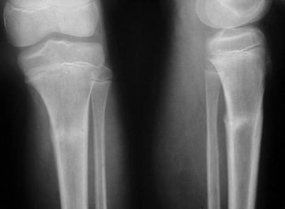
Fig. 64.2
Fracture line is clearly seen, with a small periosteal reaction
Fig. 64.3
(a, b) Stress fracture in upper end of tibia. CT scan shows periosteal reaction and changes in the density of the cortical bone
Fig. 64.4


(a, b) Stress fracture in the middle shaft of fibula. (b) The technetium bone scan shows an intense uptake over the fibular diaphysis
Related posts:


Stay updated, free articles. Join our Telegram channel
Full access? Get Clinical Tree
