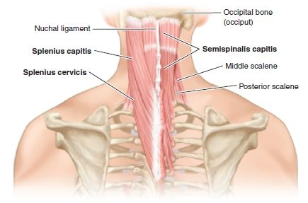
FIGURE 5.5 Posterior cervical region: Trapezius. (From Tank PW, Gest TR. Lippincott Williams & Wilkins Atlas of Anatomy. Philadelphia, PA: Lippincott Williams & Wilkins, 2009.)

FIGURE 5.6 Posterior cervical region: Semispinalis. (From Tank PW, Gest TR. Lippincott Williams & Wilkins Atlas of Anatomy. Philadelphia, PA: Lippincott Williams & Wilkins, 2009.)
PATIENT POSITION
- Sitting on an exam stool with the neck flexed and leaning forward with the arms resting on the exam table.
LANDMARKS
1. With the patient seated on the exam stool, the clinician stands directly behind the patient.
2. Locate the cervical spinous processes of the posterior neck.
3. Palpate the area of maximal tenderness in the muscles of the posterior neck. Direct pressure over this area will elicit pain and is often associated with muscle spasm. Mark the spot(s) with an ink pen.
4. At the injection site(s), press firmly on the skin with the retracted tip of a ballpoint pen. The indention(s) represents the entry point for the needle.
5. After the landmarks are identified, the patient should not move the neck.


