FIGURE 6.14 Sternoclavicular joint. (From Tank PW, Gest TR. Lippincott Williams & Wilkins Atlas of Anatomy. Philadelphia, PA: Lippincott Williams & Wilkins, 2009.)
PATIENT POSITION
- Supine on the examination table.
- The patient’s hands are folded in his or her lap with fingers interlaced.
- Rotate the patient’s head away from the side that is being injected. This minimizes anxiety and pain perception.
LANDMARKS
1. With the patient supine on the examination table, the clinician stands to the patient’s side.
2. Identify the SC joint. Palpate the clavicle in a lateral-to-medial direction. At the medial aspect of the clavicle, there is a small depression that represents the SC joint. This structure will be tender.
3. It may be helpful to rotate the ipsilateral shoulder in order to more easily identify the SC joint.
4. The injection point is directly over the SC joint.
5. At that site, press firmly on the skin with the retracted tip of a ballpoint pen. This indention represents the entry point for the needle.
6. After the landmarks are identified, the patient should not move the chest or shoulder.
ANESTHESIA
- Local anesthesia of the skin using topical vapocoolant spray.
EQUIPMENT
- 3-mL syringe
- 25-gauge, 1-in. needle
- 0.5 mL of 1% lidocaine without epinephrine
- 0.5 mL of the steroid solution (20 mg of triamcinolone acetonide)
- One alcohol prep pad
- Two povidone–iodine prep pads
- Sterile gauze pads
- Sterile adhesive bandage
TECHNIQUE
1. Prep the insertion site with alcohol followed by the povidone–iodine pads.
2. Achieve good local anesthesia by using topical vapocoolant spray.
3. Position the needle and syringe perpendicular to the skin with the needle tip directed posteriorly.
4. Using the no-touch technique, introduce the needle at the insertion site (Fig. 6.15).
5. Advance the needle into the SC joint space.
6. Inject the steroid solution as a bolus into the SC joint. The injected solution should flow smoothly into the space. If increased resistance is encountered, advance or withdraw the needle slightly before attempting further injection.
7. Following injection of the corticosteroid solution, withdraw the needle.
8. Apply a sterile adhesive bandage.
9. Instruct the patient to move his or her shoulder through its full range of motion. This movement distributes the steroid solution throughout the SC joint.
10. Reexamine the SC joint in 5 min to confirm pain relief.
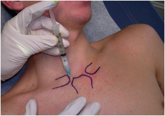
FIGURE 6.15 Sternoclavicular joint injection with landmarks.


