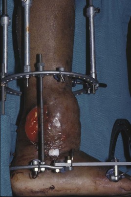
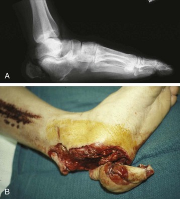
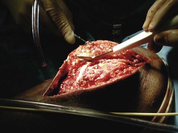
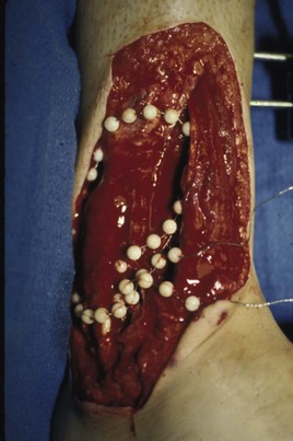
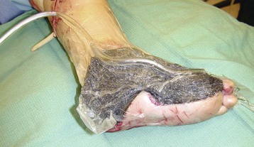
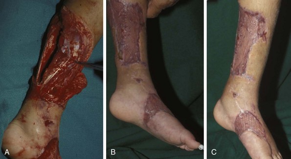
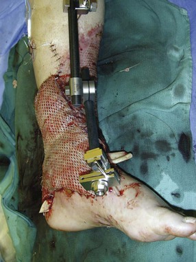
Soft Tissue Reconstruction for the Foot and Ankle

Chapter 17


Soft Tissue Reconstruction for the Foot and Ankle


Chapter Contents


HISTORY 


COMPLEX MUSCULOSKELETAL INJURIES 


OPEN FRACTURES 


THE RECONSTRUCTIVE LADDER 


DEBRIDEMENT 


SELECTION OF RECONSTRUCTION TIME 


PREPARATION OF THE WOUND FOR RECONSTRUCTION 


WOUND CLOSURE 


FLAPS 


TIMING OF FREE TISSUE TRANSFER 


SELECTION OF TISSUE FOR TRANSPLANTATION 


INFORMED CONSENT AND MEDICAL AND LEGAL ISSUES 


POSTOPERATIVE CARE 


MONITORING 


TEAM APPROACH TO EXTREMITY RECONSTRUCTION 


RECONSTRUCTIVE ALGORITHM BY REGION 


TRAUMA AND THE DYSVASCULAR AND DIABETIC FOOT 


AESTHETIC CONSIDERATIONS AFTER LIMB TRAUMA 


TISSUE EXPANSION 


History


The evolution of extremity trauma surgery reflects the development of surgery as a specialty. The management of soft tissue injuries to the foot and ankle has evolved through major historical periods that initially included high-level amputation, regional amputation to conserve limb length, wound debridement, vascular repair, flap reconstruction, revascularization, replantation, and the current concept of acute reconstruction to save the limb and preserve function.


Open fractures have plagued surgeons since the time of Hippocrates; drawings from his original writings show crude attempts at external fixation for the purpose of examining and treating wounds.2 Incomplete documents from the ancient Egyptian period reported that comminuted fractures were treated expectantly. “Compound fractures” were considered a fatal injury because amputation was not a part of the surgical arsenal.3 In the sixteenth century, Ambroise Paré, one of the founding fathers of orthopaedics, warned against the potentially life-threatening condition of gangrene resulting from open fractures; he revolutionized amputation surgery by use of the tourniquet and introduced the hemostatic clamp and vascular ligatures.68


Important advances have taken place in the field of soft tissue reconstructive surgery, including the introduction of local flaps and free flaps as efficient methods of closing large posttraumatic soft tissue defects. A better understanding of the anatomy and physiology of free flaps, with resultant high rates of success using autologous tissue transfers, and the introduction of distally based fasciocutaneous flaps have improved the surgeon’s ability to close soft tissue defects of the foot and ankle.57


With the evolution of operative methods for fracture fixation and the increasing incidence of high-energy trauma, the importance of concomitant soft tissue management in the treatment of open fractures of the foot and ankle cannot be underestimated. Foot and ankle surgeons treating patients who have traumatic conditions can no longer isolate their care to the bone or the reconstitution of articular surfaces. Although these goals are vital to the success of overall treatment, the foot and ankle surgeon must also accept responsibility for the management of the soft tissue surrounding the osseous structures and define a treatment plan for the soft tissues. This treatment plan can be carried out in conjunction with a reconstructive plastic surgeon or by the orthopaedic surgeon alone. The combination of soft tissue management and bone reconstruction permits optimal repair to take place in bone and soft tissue, avoiding the adverse sequelae of failed implants, failed fixation, sepsis, and ultimately amputation.


Complex Musculoskeletal Injuries


Complex foot injuries require effective coordinated care for both bone and soft tissue to achieve a successful fracture union while avoiding infection, but a variety of factors makes this difficult. These factors include compromised vascular supply at the fracture site, marginal soft tissue coverage, and wound contamination.


Initial treatment of fractures has improved significantly with the development of antibiotic therapy and aseptic surgical procedures in conjunction with improved stabilization techniques. This progress has increased limb salvage in the treatment of severe injuries to the foot and ankle at risk for amputation. Today’s acute fracture management with early antibiotic treatment, irrigation and debridement, and acute fracture stabilization has led to a significant reduction in fracture infection rates (Fig. 17-1).


The essential elements of osseous healing of reduced fracture fragments are blood supply and stabilization. Blood supply to bone is derived from nutrient vessels as well as surrounding muscle and fascia. Compromise of a bone’s soft tissue envelope or nutrient vasculature places that bone at risk for delayed union, nonunion, or the inability to fight infection if colonized with bacteria after injury.


Open Fractures


Historically, treatment of an open fracture in which soft tissue injuries have been neglected or mistreated has resulted in therapeutic disaster. It was argued that open reduction and internal fixation of closed or opened fractures was performed with significant risk. Arguments against internal fixation were that it resulted in infection, compromised bone healing, and nonunion.


The basic principles of open reduction and internal fixation, defined by the Arbeitgemeinschaft für Osteosynthesesfragen (AO) group five decades ago,93 included anatomic reduction and stable internal fixation, careful attention to soft tissue handling, and functional rehabilitation of the injured limb—all vital to fracture management. Functional rehabilitation involves restoring muscular power and normal biomechanics.


The importance of soft tissue reconstruction has been emphasized over the last two decades. New fixation techniques, such as indirect reduction and biologic plating with implants that respect biology and avoid compromised periosteum around bone, are now being used. Compatible implants, such as the locking plates and calcaneal plates with improved metallurgy, were designed to limit the damage to soft tissue caused by overvigorous dissection and stripping of the soft tissue envelope.85


The surrounding soft tissue has been recognized as the vascular envelope responsible for nurturing bone back to health. The importance of its reconstruction early in the posttraumatic course cannot be overemphasized; neither can the importance of surgical anatomy, internervous planes, vascular territories, and atraumatic techniques of dissection.69,115 Rather than dissection techniques that result in devascularization of bone and soft tissue, a keener awareness of delicate soft tissue handling and atraumatic technique by the orthopaedist contributes to the prevention of adverse iatrogenic sequelae after injury. Proper handling of soft tissue includes tools such as skin hooks that permit manipulation of skin and tissue flaps without further damage to the soft tissues.


To reconstruct the soft tissue envelope, the surgeon must identify which layers are deficient and the size of the deficiency. Subsequently, one must outline a treatment plan that simultaneously treats bone and soft tissue synergistically.


The soft tissue envelope is composed of several tissue layers, each with a specific function and its own vascular supply. Skin, which consists of epidermis and dermis, is the first soft tissue layer violated in the open injury, and its disruption defines the open fracture.


The next soft tissue layer, the subcutaneous tissue, is less vascular than the dermal plexus, yet it is important because it provides a cushion around bony prominences and may be quite specialized, such as on the plantar surface of the foot.


Fascia is the next deepest layer and surrounds muscle compartments. It contains a rich vascular plexus that can function as the basis for supporting tissue such as skin flaps, which can be transposed or transferred over adjacent, distant, or proximal soft tissue defects.


Muscles are richly vascularized structures that power the locomotor system. They have one of five blood supplies, as outlined by Mathes and Nahai.84 Muscles can be manipulated as transposition flaps, island pedicle flaps, and free tissue transplantations (free flaps).


The deepest layer of the soft tissue envelope is a richly vascularized layer called the periosteum. This surrounds all long bones and is vital to the response of bone to injury and repair. Recent advances in anatomic dissection and free tissue transplantation have resulted in use of the periosteum, based on the medial geniculate artery case as a free flap to augment conventional methods of bone grafting.


All of the layers of soft tissue just described, including the periosteum, will accept a split-thickness skin graft (Fig. 17-2). Despite contour irregularity, if these layers are healthy and well vascularized, a skin graft can be applied to seal the open wound. This is the first goal in wound management, that is, to reconstitute the epithelial surface of the extremity.


A logical method of reconstruction of the soft tissues must be developed to allow bone to heal and limbs to function normally. The algorithm should be capable of being used by the foot surgeon in the setting of acute or chronic soft tissue injury with or without fractures. In addition, it should be applicable to chronic conditions, such as osteomyelitis, nonunion, or tumor.26


In essence, the foot and ankle surgeon must acquire an understanding of the approach used by the plastic or reconstructive microsurgeon to reconstruct deficiencies in soft tissue. This approach, coupled with knowledge of treatment for bone deficiencies or deformities, serves as an orthoplastic philosophy of limb salvage. The reconstructive ladder represents increasingly complex solutions to correspondingly complex problems with the same goal: reconstitution of the soft tissue envelope and bone. The lowest rungs of the ladder are often as important as the highest rungs. Understanding the relationship between the needs of the wound and the various techniques offered by the reconstructive ladder is important.


The Reconstructive Ladder


Patient Evaluation


Evaluation of the patient with a soft tissue injury includes determining the time of injury, mechanism of injury, energy absorption, fracture configuration, systemic injuries, damage to the soft tissue envelope, vascularity of the extremity, sensibility, ultimate ability to salvage the foot (both functional and sensate), and underlying medical conditions of the patient. The principles of evaluation of orthopaedic trauma are the same as for any basic medical evaluation. These principles apply whether in the outpatient clinic, emergency department, or trauma unit.


An evaluation of the perfusion of the traumatized limb is of paramount importance, and if vascular (arterial) injury is suspected, a vascular surgery or microsurgery consultation should be obtained. Compartment syndrome should be considered and ruled out in any injured extremity, particularly after crush injuries. A general motor examination, including the active and passive range of motion, and a detailed sensory examination should be performed. A nerve deficit may be secondary to a spinal cord injury, nerve laceration, compartment syndrome, traction injury, or entrapment between bone fragments. The radiologic evaluation starts with a standard plain radiographic examination. Computed tomography (CT) is indicated in complex foot injuries and can give valuable information regarding soft tissue damage as well.


The wound should be inspected and the wound pattern and contamination noted. The next inspection of the wound should then be in the operating room under sterile conditions. Repetitive examination of open wounds in the emergency department has led to higher rates of wound infection and osteomyelitis and should be avoided. In cases of open fractures in polytrauma patients, workup of other injuries can take several hours, not to mention the need for emergent lifesaving visceral surgery that can precede definitive care for open fractures. Prophylactic antibiotics are administered and given on a regular basis until definitive wound debridement and fracture stabilization can be performed.


Classification of Soft Tissue Injury


A classification of the soft tissue injury should allow us to evaluate the results of the treatment, to better inform our patients, and to communicate with our colleagues in a more universal language. A classification must be reproducible and easy to remember, and it should determine prognosis of the injury. The only universally accepted classification of soft tissue injuries has been a systematic injury with division into “closed” and “open.”


Assessment of soft tissue injury is necessary in both open and closed fractures. The degree of soft tissue injury provides a prognosis and guides fracture management. The different classification schemes can be fairly simple or minutely detailed. The simpler schemes are noncomprehensive and inexact but are the most likely to be used.46


As the significance of soft tissue injuries on the influence of bone healing became more apparent, Gustilo and Anderson43 devised a three-grade classification in 1976. Type 1 fractures have a clean wound smaller than 1 cm, type 2 wounds have a laceration greater than 1 cm and without extensive soft tissue damage, and type 3 wounds are severe soft tissue lacerations with segmental or severely comminuted fractures in high-energy trauma. Because of the problems and the classification of type 3 injuries, this group was subsequently divided in three subgroups. Type 3A injury has a large soft tissue laceration or flaps but allows for adequate soft tissue coverage of bone. Also included in type 3A are fractures with severe comminution or segmental fractures, regardless of the extent of the soft tissue damage. Type 3B fractures (Fig. 17-3) are more severe, and they have extensive periosteal stripping and soft tissue loss with significant bone exposure and massive contamination. Type 3C fractures have an arterial injury requiring repair.


A more comprehensive soft tissue scale, albeit more difficult to use, is the AO soft tissue scoring system.94 It incorporates five grades of severity and three categories of tissue. The AO classification grades the skin, muscle and tendon, and neurovascular structures. Closed fractures involving only skin are graded in four subgroups. For open fractures, four grades are given. A new feature of this classification is the evaluation for muscle and tendon injuries. Because of the prognostic value, knowledge of the extent of muscle damage and tendon involvement is essential. A common approach in all classification schemes is a determination of the length of laceration of the skin. As treatment methods have become more comprehensive and more systemic factors are taken into account when treating open fractures, the presence or absence of muscle injury, nerve injury, and vascular injury has become more important prognostically. Acute systemic factors, such as shock, associated injuries, or older age, have been recognized as important prognostic indicators also. They influence the acute treatment of fractures and the treatment of complications. For example, in osteomyelitis, debilitating factors, such as smoking and malnutrition, affect the feasibility of reconstruction.12


Ruedi et al108 have developed a classification system that characterizes soft tissue injury by addressing several layers of the soft tissue envelope. This classification system determines whether the integument is open or closed. Injuries to muscle, tendon, nerve, and vessels are graded in order of severity. Although this may be more complex than the Gustilo and Anderson43 classification of open fractures, it is an attempt similar to Tscherne’s116 classification to define in more depth the deficiency and defects of the soft tissues. Factors such as contusion or ecchymosis to skin and muscle must be identified to prevent further damage to these tissues in surgical dissection. Such damage of muscle or fascia territories can make these sites unreliable as replacement tissue.


In the limb with compromised integument and a break in the epithelial surface, underlying subcutaneous tissue, muscle, fascia, bone, and periosteum are exposed, predisposing them to desiccation with inevitable cell death and the risk of infection. To help prevent infection, a sterile moist bandage should be applied to the wound as soon as possible, bathing the damaged tissues in a physiologic medium, such as saline or lactated Ringer solution.


A biologic dressing is the first step on the reconstructive soft tissue ladder. Preventing desiccation of tissue may reduce the extent of debridement and preserves simpler options for closure, such as skin grafts.


Debridement


Adequate debridement of damaged tissue is paramount in the treatment of the traumatized limb. In most instances, debridement results in additional loss of tissue, depending on the degree of contamination.


The abilities of the reconstructive surgeon, particularly the ability to transplant autogenous tissue such as muscle or skin flaps, have changed the concept of debridement.39 Surgeons treating combined injuries must accept the premise that irreversibly damaged or nonviable tissues require replacement, and the zone of injury requires expeditious reconstruction. Marginally viable tissue left behind can subsequently desiccate, infarct, and become infected, adding further delay in healing. This results in progressive dysfunction related to inflammation, fibrosis, and pain, which can be avoided if aggressive debridement is undertaken primarily. Critical vascular structures, nerves, and tendons can be cleaned, and prompt coverage can preserve their viability. Debridement may take place in the acute trauma setting or in a chronic wound that has evolved from improper handling of soft tissues.


New tools have evolved for debriding wounds. Ultrasound debridement, for example, has been used for chronic wounds. The ultrasonic methods have proved to be less painful than the conventional methods of sharp debridement when used in the outpatient clinic setting. The main advantage of the technique is that debridement can be done more precisely in areas where there are patchy areas of granulation tissue that the surgeon wants to preserve.


Other devices, such as the Versajet Hydrosurgery System (Smith & Nephew, Hull, England), enables the surgeon to cut and remove damaged tissue and contaminants while simultaneously irrigating the wound. Surgical debridement is accomplished in a single step. The Versajet (Fig. 17-4) uses a high-velocity stream of sterile saline that jets across the hand piece into a vacuation collector. The Versajet requires less irrigant than traditional techniques, and it confines the irrigant to the wound area. In the acute trauma situation, this obviates the need to change large saline irrigant bags and reservoir waste canisters. This system has several power settings that can be used depending on the degree of debridement needed. One advantage of the hydrosurgery system is that there is less aerosolization of bacteria, which decreases risk to the operating room staff and surgeon based on a single wound site. Compared with conventional pulsate irrigation, the Versajet leaves significantly less bacteria in the wound.


Fresh Wounds


Debridement of the fresh wound (such as an open IIIB or IIIC pilon fracture) should be performed in an exsanguinated extremity. This permits the surgeon to carefully observe the appearance of the tissue and detect pockets in the wound, and it eliminates all foreign material. In addition, operating under tourniquet prevents unnecessary blood loss during debridement. After debridement is completed, it is relatively easy to identify major bleeders and perform proper hemostasis once the tourniquet is deflated. After release of the tourniquet, the result of debridement can also be checked by observing diffuse bleeding throughout the wound.


In the ischemic operative field, it is relatively easy to distinguish between healthy and damaged tissue. The basic elements of this judgment are the appearance and consistency of tissues. Healthy tissue in the exsanguinated limb is bright and homogeneous in color. Subcutaneous tissue is yellow, muscles are bright red, and the tendons and fascia are white and shiny. Damaged tissues are recognized by the presence of foreign bodies, irregular tissue consistency, and irregular distribution of dark red stains, which are hematomas.


All nonviable tissue is removed, preferably with a knife. It is not possible to perform exact debridement with scissors. Scissors should be used only when dissecting important structures such as nerves, vessels, or tendons.


The edge of the debridement should be in healthy tissue. Avulsed skin and muscles should be removed from the base of the avulsed flap. These tissues are contused; the anatomic pattern of the skin’s vascularity is not “axial” because of avulsion of perforators, and the vascularity is therefore insufficient to maintain viability of such flaps. Denuded tendon, if not frayed, should be cleaned. Disrupted tendons can be sutured or can be fixed with a suture to surrounding tissue for later reconstruction. Exposed bones are washed with antibiotic solution and mechanically cleaned with bone rongeurs. Free bone fragments are usually removed, and if sections are quite large, they can be stored in the bone bank as bone autografts for later use when soft tissue coverage has been obtained. However, this point is controversial.


Severed vessels are ligated, provided they are not critical to the viability of the injured extremity. If vessels are vital, they are excised to normal-looking margins, and continuity is restored with interposition vein grafts.


The nerves are the only structures where debridement is not radical. Those parts that are destroyed without any doubt are removed, and major nerve stumps are anchored in the wound to prevent retraction and allow later reconstruction with nerve grafts. We prefer that a large cystoclip or hemoclip be applied to the nerve stump so it can be seen on a radiograph and appropriate secondary planning can be done in terms of surgical approach and location of the nerve stump.


Occasionally, the bed of the wound remains irregular after debridement. With additional incision, it can be made regular in shape, provided that the irregularity is not caused by tissue of functional importance. This maneuver serves to eliminate dead space by permitting the flaps, if they are to be used, close contact with the wound bed, preventing hematoma and subsequent formation of scar.


Chronic Wounds


Debridement of a chronic wound is also done with the limb exsanguinated. Superficial scar is excised completely so that the wound margins are in healthy skin and subcutaneous tissue appears normal. The goal is to treat the chronic wound like a tumor and excise it in its entirety down to normal tissue planes. All scars in the wound should be removed in the same manner as cancer surgeons operate; that is, the knife should always cut through healthy tissues. If functionally important structures are entrapped in the scar, the dissection should commence in the healthy surrounding tissue and pass toward the scar entrapment, where the structures such as nerves or tendons are carefully dissected and preserved.


This portion of the procedure is extremely difficult, and it is necessary to anticipate the changes in the anatomy caused by the injury, previous operations, and the traction exerted by scar tissue. Bone debridement is also very difficult. Although clearly necrotic parts of bone can be recognized, more experience is required to identify the viable parts of the bone callus and necrotic and inflamed areas in bone. Studies such as CT scans, bone scans, and magnetic resonance imaging (MRI) may be helpful in preoperative planning for bone and soft tissue debridement.


Swiontkowski114 has popularized the use of the laser Doppler to determine bone blood flow in planning for debridement of infected bone. All areas of the bone not covered with periosteum are removed, and those that are exposed are burred with an iced saline bur. If punctate bleeding is encountered from the cortical bone, the bone is left behind. If not, the bone is removed until the paprika sign is identified. That is a sign of bleeding bone and live bone. This is punctate bleeding from the haversian canals that indicates bone viability. If a sequestrum is in the medullary canal, the anterior part of the bone cortex (such as the metatarsal) should be removed to provide a window for access to the medullary canal for placement of muscle flaps, which eliminates dead space and helps control infection.


Debridement Technique


Surgical Technique


1. The extremity is prescrubbed to remove grime and surface dirt, and then it is prepared and draped. Nails should be cut and nail plates cleaned.


2. The leg is elevated for 5 minutes to exsanguinate it (rather than wrapping the extremity with an Esmarch bandage), and the tourniquet is inflated.


3. The wound is superficially washed with an antibiotic-saline solution to remove blood clots and superficial debris. It is advisable to use loupe magnification when debriding. The more complex the wound, the longer the debridement takes. If the patient with an open wound has been waiting for quite some time and blood is organized in muscle tissues or around fracture ends. Half-strength peroxide can be used as a first rinse solution to lyse the clot and gain access to the true depths of the wound. Half-strength peroxide has a tendency to bubble and should be washed away with normal saline solution.


4. A No. 10 or 15 scalpel blade or very sharp scissors is used to excise the skin and dermis, particularly around the edge of the wound, back to normal tissue.


5. The subcutaneous layer is inspected and debrided sharply with a No. 15 scalpel blade to the level of fascia. All fascia that is stripped, avulsed, or contaminated should be removed.


6. The next layer encountered is muscle. Muscle should be resected down to healthy tissue, regardless of the amount of muscle removed. Leaving unhealthy necrotic muscle is one of the surest ways to initiate an infection.


7. Periosteum that is elevated from bone should be excised to the level from which it is elevated. Small bone fragments devoid of periosteum or free-floating large segments, even structural ones, should be removed for fear of colonization, contamination, and infection.


8. At the conclusion of debridement, the wound is again irrigated. The tourniquet is released and all tissue planes, particularly the muscle, are observed for bleeding as the arterial pressure increases within the limb.


9. Areas that remain nonviable, particularly the dermis, skin, and muscle, are reexcised. Excision then can be done sequentially, watching for punctate bleeding from the dermis or the muscle. When large flaps have been avulsed, excision is carried out through the skin to the level of bright red blood coming from dermis on incision.


10. No attempt should be made to close the wound defect under any tension, for fear of further ischemic damage to already compromised tissue.


Special Techniques in Debridement


In the chronic bone infection, such as in the infected calcaneus, a motorized burr may be used to debride bone. Bone that is easily removed by the burr is necrotic and nonviable. At the point where there is punctate bleeding from the cortical and/or cancellous surface, indicating good viability of bone, the burring is stopped. Pulsed irrigation systems, although advocated by some, if used too vigorously with too high pressure too close to tissue planes, particularly around tendons, nerves, and vessels, can actually damage tissue and cause swelling. For this reason, we advocate copious but gentle irrigation. A wound that is appropriately debrided sharply down to all healthy tissue requires very little irrigation.


The philosophy of wound debridement should be similar to tumor resection: visualizing all normal tissue planes at the conclusion of debridement. If this is not possible, then we advocate a second-look procedure in which the debridement process is repeated, particularly if there is questionable viability of tissue. This should be done no later than 48 hours, and preferably 24 hours, after the initial debridement. If possible, during the first and not later than the second debridement, plans should be made to obtain wound coverage by one of several methods.


Within the reconstructive ladder, options are now available to reconstruct bone as well as soft tissue. No compromised tissue should be retained. The “wait and see” attitude concerning bone devoid of periosteum or muscle that is not bleeding but is covering a vital structure should be abandoned. Adequate surgical exposure is critical in the assessment of soft tissue injury as well as in its treatment.


Timing of Reconstruction


The optimal time for soft tissue reconstruction in severe open fractures remains controversial. The argument favoring a staged method is the need for a second-look debridement. If there is uncertainty about traumatized and devascularized tissue, a second look is performed. The main argument for early reconstruction is to reduce the nosocomial contamination and secondary necrosis of exposed tissues. Late soft tissue reconstruction is associated with a significantly higher infection rate and flap complication rate when compared with early (within 72 hours) soft tissue coverage.20,36


Godina40 and other pioneers64 changed the concept of primary repair and reconstruction of damaged tissue by advancing the phase of reconstruction from a delayed elective procedure to the day of injury. Assuming that an adequate primary debridement is feasible, the outcome should be improved by immediate soft tissue closure to avoid bone infection. Immediate reconstruction improves the time to definitive fracture union, decreases the number of operations that are performed, and reduces infection rates.56


Patients with IIIB and IIIC open fractures of the foot and ankle whose general condition was suitable for debridement, followed by stable internal fixation and immediate soft tissue reconstruction, demonstrated a better outcome and a shorter period of convalescence.56 The higher incidence of infection in the delayed group may well be due to the lengthy exposure of the fracture to nosocomial contamination, the secondary damage of exposed tissue, or the necessarily incomplete nature of second-look debridement, particularly in and around a reduced fracture.


Preparation of the Wound before Reconstruction


Wound Dressing


The standard protocol for wound management associated with fractures after surgical debridement varies with the severity of the wound. In grades I to III open fracture wounds, the gold standard technique before definitive wound coverage has been to pack the wound with saline-soaked gauze dressings, which helps eliminate dead space and prevents soft tissue desiccation. The disadvantages of saline-soaked gauze dressings include drying with soft tissue desiccation, nosocomial bacterial contamination, poor dead space management, and, often, significant patient discomfort. Similar benefits are obtained with dressings of gauze soaked in Dakin solution and half-strength povidone-iodine (Betadine). Dakin solution is bacteriostatic. Povidone-iodine, although bacteriocidal, is controversial because it is toxic to soft tissue. The advantage of all three of these dressing types is that they ensure cleanliness at the time of closure by allowing consistent monitoring of the wound site.


Fracture wounds with avulsion of the dermal surface but without damage to the underlying muscle may be treated successfully with several techniques. First is emollient coverage. Emollient-type soft tissue coverage may also be indicated to temporize a wound before soft tissue coverage. This may take the form of a hydrogel such as Vigilon (Bard Medical, Covington, Ga.), an antibiotic-impregnated occlusive dressing such as Scarlet Red (Kendall Healthcare, Mansfield, Mass.), or a simple semipermeable film such as OpSite (Smith & Nephew, Memphis, Tenn.). A copious layer of antibiotic ointment covered by a Vaseline dressing and gauze can also temporize wounds before soft tissue coverage.


There are four general types of newer wound dressings: semipermeable films (e.g., OpSite, Tegaderm [3M, St. Paul, Minn.], and bio-occlusive), hydrogels (e.g., Vigilon), occlusive hydrocolloids (e.g., Duoderm, Synthes, West Chester, Pa.), and synthetic skin substitutes (e.g., Epigard [Synthes, West Chester, Pa.] or Integra [Integra LifeSciences Corp., Plainsboro, N.J.]).34 These newer dressings are best applied when the wound site is surrounded by a border of healthy tissue.


Semipermeable films and semiocclusive hydrogels are impermeable to water and bacteria but permeable to oxygen and water vapor. Occlusive hydrocolloids are impermeable to even water vapor and oxygen. For example, Duoderm has an inner adherent surface with an outer impermeable polyurethane foam. Epigard, one of the synthetic skin substitutes, is a nontextile open matrix polyurethane composed of two layers and backed by a microporous polytetrafluoroethylene (Teflon) film. This matrix allows new microcirculation to develop in its interstices.


Newer dressings are not without drawbacks, however, particularly the accumulation of exudate, hematoma, and seroma beneath them.34 In addition to these wound dressings, there is an isolated report on the efficacy of honey as a broad-spectrum antimicrobial found to be effective in controlling Staphylococcus aureus, Escherichia coli, Pseudomonas aeruginosa, and Klebsiella pneumoniae. Although this finding needs further clinical verification, honey shows promise for use as a first-line wound dressing agent.52


Antibiotic Beads


Antibiotic beads have been used effectively to prevent or to control infection and can be used after the first debridement until the patient returns to the operating room for a second-look debridement (Fig. 17-5). Advantages of antibiotic beads are that they can deliver antibiotics at high concentration to a compromised wound without systemic effects. The bead pouch, popularized by Henry et al,55 seals the wound so that transudate from the wound surface is captured, bathing exposed tissues in physiologic fluids that also contain bactericidal levels of antibiotics. Wounds do not desiccate, cell death is avoided, and infection risk is reduced.


Antibiotic Bead Pouch

The technique of local antibiotic therapy achieved via antibiotic-impregnated polymethyl methacrylate (PMMA)54 originated with Büchholz in Germany. It was an application that used antibiotic-impregnated bone cement to treat infected arthroplasties.53


Antibiotic-impregnated PMMA beads are strung on steel surgical wire. A chain of medium-sized 6.3-mm PMMA beads is composed of 21 beads, each bead weighing 70 mg and containing a 5.7-mg tobramycin base. A chain of smaller beads has 20 beads that are 2 mm in diameter, each weighing 14.5 mg with a 2.2-mg tobramycin base. The surgical wire consists of three strands of size 2-0 surgical wire for the 6.3-mm beads. With the smaller size, four strands of 4-0 steel sutures are used.123 The custom-made versus commercial fabrication processes differ in that the custom-made beads are polymerized in a mold, whereas the commercial variety is formed in a press with a corresponding increase in temperature and pressure.


The bead pouch technique is most effective for grade III open fractures. It is indicated for use in grade I fractures only if primary closure was prevented because of compartment syndrome, marked swelling, or wound edema.54 The bead pouch technique is performed in the operating room under sterile conditions.


Surgical Technique


1. All necrotic, avascular, and contaminated tissue is removed from the fracture site during the initial irrigation and debridement. Wound margins are extended to appropriate widths.


2. A thorough lavage consisting of bacitracin and normal saline is performed.117


3. Depending on the severity of the fracture, reduction is accomplished with either external or internal fixation.


4. One or more chains of antibiotic beads are inserted into the wound surrounding the fracture site. Placement should be such as to fill the soft tissue cavity but leave adequate room for closure.


5. A suction drain (0.32-cm diameter) is placed in the wound. The drain should be positioned so that it exits the hematoma site through normal tissue.117


6. If possible, the wound should be closed with interrupted sutures. (In wounds with extensive soft tissue damage, closure may not be possible at the time of initial debridement.)


7. Wound coverage is achieved with an adhesive polyethylene wound film, such as OpSite. The semipermeable wound dressing should be stapled to the skin edges and a second layer wrapped around the entire wound area to prevent leakage of wound secretions.


8. The drain should remain in place for 48 hours. Suction is avoided because it would negate the high bactericidal dosages released by the beads into the wound hematoma.


9. The bead pouch is replaced every 48 to 72 hours in the operating room under sterile conditions. This is done to ensure adequate antibiotic concentrations in the wound environment. Aerobic and anaerobic cultures are taken at each bead change. Final wound closure is achieved through either primary suture closure or, in cases with more extensive soft tissue defects, split-thickness skin grafts or flap coverage.


Discussion

In more severe fractures, the bead pouch provides a solution to much of the debate surrounding delayed or acute soft tissue transfer. Because the bead pouch delivers high levels of antibiotics locally, the surgeon can delay definitive coverage until thorough and usually multiple debridements have been performed, a clean wound is achieved, and operative repairs of neurovascular, tendon, and ligamentous structures are made.54 If an acute flap is indicated, the presence of the bead pouch beneath the flap serves to assuage the fears the surgeon might have that infection could occur beneath the flap.


The bead pouch technique shows significant advantages over saline-soaked gauze dressings in preventing desiccation and subsequent soft tissue necrosis. The bead pouch provides an environment in which the bone is enveloped in a moist, protective envelope. This allows for greater infection resistance, increased wound vascularization, and a therapeutic level of antibiotics to be administered at the wound site.


Hyperbaric Oxygen


Hyperbaric oxygen (HBO) can be used to promote formation of granulation tissue and stimulate angiogenesis in wounds that are compromised, usually by impaired arterial inflow or venous outflow.66 In addition to exposure to hyperbaric oxygen, wound dressings may be changed under sterile conditions by chamber personnel under sedation, avoiding discomfort to the patient. Patients who have gas gangrene associated with fractures require emergent debridement, hyperbaric oxygen, antibiotics, and ultimately fracture and soft tissue management.91


Normal tissue oxygen levels are approximately 40 mm Hg. When tissue levels fall below 30 mm Hg, normal metabolic activity is significantly impaired.42 In infected wounds and traumatized tissue, oxygen levels often fall to less than 30 mm Hg. HBO enhances oxygen delivery to ischemic and hypoxic wounds, and even when it causes local vasoconstriction, the overall increase in blood oxygen content results in a net gain so that the net oxygen concentration at the wound increases. HBO improves neutrophil function, facilitates fibroblast cell division, increases collagen formation, and encourages new capillary budding. The promotion of angiogenesis by HBO is thought to be one of the major factors in promoting the healing of chronic hypoperfused wounds.59


Negative Pressure Therapy


Negative pressure therapy (NPT) exposes a wound to subatmospheric pressure. It has proved to be extremely effective in treating a wide spectrum of wounds, including traumatic wounds as well as dehisced incisions with or without exposed hardware.6


The wound cavity is dressed with a cell foam dressing that is connected to an adjustable vacuum source with a negative pressure of up to 125 mm Hg. The foam dressing and wound site are sealed with a thin adhesive film, converting the open wound to a controlled closed wound. Pressure is applied continuously or cyclically to the wound (Fig. 17-6). The removal of excess interstitial fluid from the wound periphery results in a decrease in the local interstitial pressure, thus restoring blood flow to compressed or collapsed vessels. Along with removal of the chronic fluid, factors that inhibit healing are also removed. An additional mechanism of action of NPT is the mechanical stimulation of cells by tensile forces placed on the tissue because of the collapse of the foam dressing by the negative pressure.


NPT closure has become an important adjunct of foot and ankle care. This device decreases edema, increases wound vascularization, fights infection, and promotes granulation tissue through proliferation of capillaries and fibroblasts.


NPT has several applications in foot and ankle surgery. It can be used for soft tissue injuries after initial debridement. If a relatively clean wound can be established, a wound vacuum-assisted closure can be applied. This device serves as a barrier dressing, and it isolates the wound from the hospital environment. Simultaneously, it encompasses all of the physiologic benefits that have been mentioned. It establishes an environment for granulation to occur.


After a second-look procedure, NPT may be used as definitive wound care treatment, usually for large cavitary wounds or wounds with irregular surface topography; a wound NPT can also stabilize the wound until definitive soft tissue treatment can occur. For example, an open calcaneus fracture that has a large cavitary soft tissue component can benefit from a wound NPT to serve as a barrier dressing from the hospital environment. When the patient is taken back to the operating room, NPT can be removed and appropriate soft tissue reconstruction can be performed with skin grafts or, more commonly, with free tissue transfer.


The advantage of NPT is similar to that of an antibiotic bead pouch in that the wound is sealed from the outside. The advantage of the bead pouch is that there is a high concentration of antibiotic that can be released into the tissue. The wound NPT relies more on physiologic rather than pharmaceutical treatment of the wound in that blood flow can be enhanced and edema can be diminished, making the wound healthier, assuming parenteral antibiotics are given.


Another application of the wound NPT has been in the treatment of fasciotomy sites. Traditionally, fasciotomy sites are covered with materials such as biosynthetic skin substitute (Biobrane, Smith & Nephew, Memphis, Tenn.) or saline dressings that require frequent dressing changes to prevent wound desiccation. In an era when hospitals are trying to diminish global costs, particularly in the trauma population, the wound NPT can remain on a wound for 2 or 3 days, and sometimes longer, depending on the clinical circumstances. For example, the wound NPT has been a very effective stabilizer of skin grafts that are used for definitive coverage of open wounds or be skin grafted to fasciotomy sites. In those cases, the wound NPT stays on for approximately 5 days with a pressure of −75 mm Hg.


Although it is possible to definitively treat certain wounds to completion with a wound NPT, meaning complete epithelialization, a word of caution should be added regarding the use of wound NPTs with exposed hardware, such as fixation plates and screws. If the NPT is applied long enough, it is possible that a wound will granulate such that the granulation grows over exposed plates and screws. The granulation tissue contains bacteria that can colonize the plates and screws, resulting in chronic infection. In addition, the ultimate healing of soft tissue using NPT creates a healed wound, and the wound has a large degree of fibrosis associated with it. If one needed to reoperate on a foot that was treated with NPT and a skin graft, compared with supple soft tissue such as a muscle flap or regional transpositional flap, working through the scarred bed would be difficult, resulting in complications after secondary procedures, such as tendon transfers, bone grafting, or hardware exchange or removal.


Amputation versus Salvage


In complex extremity injuries, the treating physician must first determine whether limb salvage is feasible. Before complex and prolonged reconstructions are started on a limb that will ultimately function poorly or not at all, a well-fitted prosthesis should be seen as an excellent therapeutic option, and early amputation should be considered. Lange73 and Hansen47 have delineated a sound algorithm for these difficult cases.


If neurovascular structures are injured, are they repairable? Is normal plantar sensation possible? Does a compartment syndrome exist? Unless a compartment syndrome is recognized and treated, muscle ischemia and death will occur, converting potentially viable soft tissue to infarcted muscle and scar. This ultimately increases the need for large bloc resections and tissue replacement—a higher rung on the reconstructive ladder.


Although the evolution of sophisticated microsurgical reconstructive techniques has created the possibility of successful limb salvage in even the most extreme cases, such technical possibilities are double-edged swords. Hansen,47 in analyzing his vast personal experience with managing open fractures, noted that protracted limb salvage attempts might destroy a patient physically, psychologically, socially, and financially, with adverse consequences for the patient’s family as well. In spite of the best attempts, the functional results of limb salvage are often worse than those of an amputation. Thus enthusiasm for limb-salvage techniques, especially of the traumatized foot and ankle, must be tempered by a realistic assessment of the results, not just for the injured part but for the patient as a whole.73 A salvaged limb must function as well as, if not better than, a prosthesis, or heroic attempts at reconstruction are not indicated. Donor-site morbidity should also be considered with free tissue transfer when considering limb reconstruction.


Indicators of a poor prognosis for limb salvage, in order of significance, are massive crush injuries, other high-energy soft tissue injuries, a warm ischemia time longer than 6 hours, severely comminuted or segmental fracture patterns, infrapopliteal arterial injury, prolonged severe hypovolemic shock, and age older than 50 years.


Occupational demands and the presence or absence of underlying diseases, such as diabetes, are also important considerations. The same anatomic injury can require a different treatment decision in a 20-year-old laborer than in a 60-year-old diabetic patient.


Similarly, a given tibial injury might need to be treated differently if there is unreconstructible ipsilateral foot trauma that precludes reasonable limb function even if the leg is salvageable. According to several authors, complete disruption of the posterior tibial nerve (in association with type IIIC tibial fracture) is also a functional liability significant enough to warrant amputation.21,58,74 Decreased sensation on presentation is not an indication for amputation.


Expedient amputation of a massively traumatized limb, even if it appears salvageable, may also be necessary in the multiply injured patient who cannot tolerate the reconstructive time or metabolic demands of the reconstruction. This is an extremely difficult judgment that is highly individualized and impossible to quantitate.77


Wound Closure


After adequate debridement, it may be possible to close a wound primarily. This is done rarely, if ever, in cases of open fracture resulting from the danger of contamination or the need for a second-look debridement. However, approach incisions can be primarily closed after extensive aproaches for debridement.


The second rung in the reconstructive ladder is delayed primary closure. This method is considered in cases in which the adequacy of the primary debridement is uncertain; it is usually done after the initial edema subsides. Healing by second intention refers to epithelialization and wound contraction. This technique may also be applied to abrasions, avoiding the need for skin grafts.


The next level of reconstruction is use of a split-thickness skin graft (STSG), either meshed or unmeshed.


Skin Grafting


The skin is composed of dermis and epidermis. The dermis contains sebaceous glands, most of which are appendages of hair follicles.107 Sweat glands and hair follicles are mostly located in the deep dermis. These skin appendages are lined with epithelium, thus allowing for re-epithelialization after removal of epidermis and partial removal of dermis, as in superficial burns or harvesting of split-thickness skin.


Although sebaceous glands are not under hormonal control and continue to function after skin grafting, sweat glands (both apocrine and eccrine) temporarily lose nervous control. As a result, newly grafted skin may be dry, requiring moisturizing with common over-the-counter preparations.


What type of wound can be healed with a skin graft? Any wound with a full-thickness loss of skin may be considered for skin grafting. Such defects might heal spontaneously but, because of the lack of dermal appendages, do so through the process of contraction and epithelialization from the wound periphery. When healed, these wounds have skin lacking the normal microanatomy found in native skin. Specifically, there are no rete pegs—epidermal projections into the dermis that anchor the two layers of skin together. Such a wound is relatively unstable in terms of long-term durability. Allowing spontaneous healing of a full-thickness wound is therefore only appropriate for small defects and for certain larger defects where the contraction process will not distort critical anatomy or cause a functional disturbance, such as a joint contracture.


In evaluating a wound for possible skin grafting, the surgeon must assess the adequacy of the wound bed. The process of graft healing will require ingrowth of vessels from the wound, and so relatively avascular tissue provides a poor bed for skin grafting. Exposed bone, tendon, nerve and cartilage, necrotic tissue, and devascularized fat are examples of tissues that provide a poor wound bed. Conversely, well-vascularized tissues, such as muscle, periosteum, fascia, healthy fat, and epitenon, will all accept skin grafts.


Split-thickness skin grafts (STSGs) contain less dermis than full-thickness skin grafts (FTSGs), thus allowing more of the natural wound-healing process of contraction to occur. They also have less metabolic demand relative to FTSGs and so are more likely to succeed on a vascularly compromised wound. FTSGs are thicker; more durable in the long term; can carry hair follicles, allowing future hair growth; contract less; and retain more of their natural donor-area pigmentation. In general, FTSGs retain greater sensation than do STSGs.


Because one can expect epidermis to regenerate after an STSG is harvested, donor sites can be large. Despite the spontaneous regeneration noted in these donor sites, however, there is still a permanent scar, usually noted by subtle textural changes. Therefore, in certain ethnic groups with a propensity for hypertrophic scarring, strict attention must be paid to where split-thickness skin is harvested. In some Asian or African American patients, for example, scalp skin may be preferable to thigh skin for this reason. The most common donor sites for STSGs, in order of cosmetic preference, are buttock and thigh, abdomen, back, scalp, chest wall, and arms.


Harvesting of an FTSG requires closure of the donor wound because spontaneous re-epithelialization is not possible. As a rule, FTSGs tend to be small and are usually used for reconstruction in the foot.


The healing of skin grafts has been described as a two-step process. Initially, the graft survives by imbibing wound-bed exudate. After approximately 48 hours, new blood vessels begin to sprout from the wound bed and grow up into the graft. Either capillaries from the wound bed connect with graft vessels, a process known as inosculation, or they form new vascular channels within the graft—revascularization. During these processes, fibrin and collagen are also assisting in forming a cohesive bond between graft and bed. By the fifth to seventh postgraft day, a skin graft is usually adherent and vascularized.


Split-Thickness Skin Grafting

Before skin grafting, the wound must be adequately prepared. All nonviable material must be removed sharply so that the wound bed is left with healthy, preferably bleeding, tissue. A curette is quite useful in this regard. Adequate hemostasis is imperative; however, overuse of electricocautery leaves a charred wound that is unlikely to foster inosculation and revascularization of the graft. Most often after debridement, the wound is elevated and covered with a sponge soaked in normal saline. By the time the graft has been harvested, the wound bed is usually quite dry. Topical hemostatic agents, such as thrombin, may also be used.


Equipment needed to perform an STSG consists of a dermatome, mesher, mineral oil, suture or staples, and dressings appropriate for both graft and donor site. Dermatomes have evolved from hand-held knives (e.g., Humby knife) to electric and gas-powered devices able to precisely harvest skin of various thickness. Most dermatomes require insertion of a disposable blade and have two adjustments that need to be made by the surgeon: thickness and width.


Thickness, measured in thousandths of an inch, is usually set with a dial on the dermatome itself. For most patients, graft thickness is between 0.012 and 0.017 inch. The clinical setting dictates an appropriate thickness. One must balance survival (short and long-term) of the graft with donor-site healing. Therefore, in an elderly patient with thin skin, a thinner STSG (0.012-0.013 inch) may be chosen, whereas in a younger patient a thicker STSG (0.015-0.016 inch) may be preferable. The surgeon must also choose an appropriate width of skin to harvest. This adjustment is either on the dermatome itself or dependent on an attached cover placed over the blade before harvesting.


Surgical Technique


1. The donor site is prepared by first removing any povidone-iodine and other foreign materials from the skin. Mineral oil is applied liberally to both skin and dermatome to aid in gliding.


2. The dermatome is turned on, placed at a 45-degree angle with the skin, and advanced firmly to prevent slippage. As the graft is being harvested, punctate bleeding from a white dermal bed should be seen. If subcutaneous fat is noted, harvesting should be stopped immediately, the graft replaced, a new donor site chosen, and the thickness setting on the dermatome readjusted.


3. After an adequate amount of skin has been harvested, the dermatome is lifted from the donor area.


4. The donor area is temporarily covered with a hemostatic agent (local anesthesia with epinephrine or topical thrombin is a popular choice). (Donor-site dressings are reviewed later in this discussion.)


5. The skin graft is then meshed. Meshing is necessary for two reasons. First, STSG failure is usually secondary to hematoma formation beneath it, and meshing of the skin allows drainage postoperatively. Second, when a large amount of graft is needed (e.g., for a large burn), meshing also allows the graft to expand so that a relatively small piece of skin can cover a larger area. Most popular meshers require placement of the skin on a “carrier” chosen for the ratio of mesh. Unless a paucity of STSG donor sites requires a larger ratio, most grafts are meshed at 1 to 1.5. Disadvantages of meshing mostly relate to the aesthetics of the healed meshed skin, which retains its “meshed” appearance in the long term (Fig. 17-7).


Stay updated, free articles. Join our Telegram channel

Aug 27, 2016 | Posted by in ORTHOPEDIC | Comments Off on Soft Tissue Reconstruction for the Foot and Ankle

Full access? Get Clinical Tree

Get Clinical Tree app for offline access