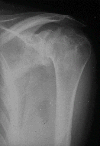
Fig. 5.1
Shoulder AP radiograph of a patient with pain and bleed episodes who has developed moderate degenerative changes with cysts in the humeral head and glenoid

Fig. 5.2
Shoulder AP radiograph of a hemophilic patient with pain. There is significant elevation of the humeral head secondary to a massive chronic cuff tear and numerous cysts in the humeral head
In the same study, radiographic changes that correlated with the patient’s clinic were observed. Only 32 % of patients with mild changes presented symptoms, while in those with moderate or severe changes, the percentage increased to 59 and 65 %, respectively.
The use of MRI in hemophilic arthropathy of the shoulder is very useful in the examination of soft tissues. With its use, the synovium bursa, tendon sheath, cartilage, and rotator cuff can be evaluated. Ultrasonography has proved a viable alternative on occasions in which an MRI cannot be conducted or is unavailable. Both methods have comparable precision in the identification of partial and total tears of the rotator cuff [5]. Ultrasonography is also a useful tool in identifying hemarthrosis and choosing the most appropriate treatment [1].
Patients with shoulder involvement most of the times require a TC before surgery to identify bone deficiencies, especially if a joint replacement is being considered. TC allows evaluation of the glenoid bone stock which would help in deciding if a total shoulder arthroplasty or a hemiarthroplasty is preferred [4].
5.4 Management
The key to the successful prevention of hemophilic arthropathy is management of initial hemarthrosis before the development of chronic synovitis and articular surface erosions.
As a result, prophylaxis studies have been developed. There is a distinction between primary and secondary prophylaxis. Primary prophylaxis would be used after the initial diagnosis or after the first bleeding episode, maintaining levels of clotting factor between 1 and 5 % via repeated injections [6]. However, this would be very expensive. The alternative treatment is secondary prophylaxis consisting of clotting factor injections after every bleeding episode until the complete recovery of acute hemarthrosis occurs [7].
5.4.1 Acute Hemarthrosis
In cases of acute hemarthrosis, the priority is to distinguish between patients with established arthropathy and those without. To that end, it is useful to use ultrasonography to discern the degree of hemorrhage and joint degeneration.
In patients with established arthropathy, the only reason to conduct an arthrocentesis would be to relieve pain and possibly improve mobility.
In cases in which there is no damage to the joint, arthrocentesis is recommended after factor replacement to avoid the buildup of proteolytic enzymes, immobilizing the joint for 2 weeks, followed by 2–4 weeks of physical therapy.
Steroid injection, combined with immobilization, is effective in some patients with chronic hemarthrosis [4].
5.4.2 Synovectomy
Synovectomy is recommended in cases of chronic synovitis in which no joint degeneration has occurred. While synovectomy prevents bleeds, thereby delaying joint degeneration, the procedure does not reverse changes that have already taken place in the joint.
There are documented gains from synovectomy. First, analgesia gained for excision the inflamed tissue. Secondly, the number of bleeding episodes can be reduced, and with this, the possible delay of the hemophilic arthropathy [8].
There are three methods for performing a synovectomy: medical (synoviorthesis), arthroscopic, and open.
Medical Synovectomy (Synoviorthesis)
This method acts as a chemical synovectomy and prevents synovial proliferation by introducing a fibrosing agent inside the joint that strangles the vessels.
Although many materials are available, radiocolloids have been the most widely used. Radiocolloids applied in synoviorthesis include gold (Au 198 – no longer used due to gamma radiation), rhenium (Re 89), and yttrium (90 Y). The advantage of using these materials is that they require only one dose. The disadvantage is the high cost.
Rifampicin and oxytetracycline chlorhydrate are being examined as inexpensive alternative products. They both have similar fibrosing action and are used in a similar fashion in pleurodesis [9].
In the study by Fernández-Palazzi et al., 82 patients who had received repeated oxytetracycline chlorhydrate injections in the knees, elbows, and ankles were examined retrospectively. Significant improvements were observed in both range of mobility and pain [9].
Rezazadeh et al. studied the use of rifampicin injected into the knees, elbows, and ankles of 21 patients, and in the shoulder of one patient. A mean reduction of 6.3 bleeding episodes per month was obtained [10].
In conclusion, this method may reduce hemarthrosis, related pain, and also improve the range of motion in patients with hemophilic arthropathy. Chemical synoviorthesis appears to be efficient. The use of rifampicin and oxytetracycline chlorhydrate offers an inexpensive and simple alternative that may prove especially practical in developing countries where radioactive agents are not easily available. The disadvantage is that it may be necessary to repeat injections several times.
Arthroscopic Synovectomy
The primary indication for arthroscopic synovectomy is recurrent joint hemarthrosis with failure of appropriate medical management. Secondary indications include joint pain and loss of motion. The primary contraindication is advanced degenerative joint disease.
Stay updated, free articles. Join our Telegram channel

Full access? Get Clinical Tree


