The shoulder and elbow represent two of the most commonly injured joints in the adolescent population. Specific injuries vary by sport and can involve various structures, depending on the mechanism of injury. Unlike the adult shoulder, the immature skeletal structure of the adolescent athlete can lead to several unique injuries. By understanding the special demands placed on the immature shoulder, the sports physician can more effectively treat the resultant injury. This article reviews the diagnosis and management of unique injuries to the shoulder and elbow in the adolescent athlete.
Shoulder and elbow pain in the adolescent population is different than in the adult population. The difference relates to the immature bone structure of the shoulder and elbow region. although beyond the scope of this article, it is important to understand the maturation of the physis or growth plates. In brief, the physis or epiphyseal plate is where future bone is laid down and is constantly changing during the years of growth. The relative weakness at the physis and decreased resistance to shear and tensile forces compared with the surrounding ligaments, tendons, and muscles predispose this area to potential injury . It is important to take into account maturation of the bone, including the closing of ossification centers, when evaluating the immature athlete. Important ages to remember for closure of the ossifications centers of the shoulder and elbow region include the upper part of the glenoid (16–18 years), the proximal humerus (17–18 years), the lateral epicondyle (14–16 years), and the medial epicondyle (14–16 years).
Specific shoulder and elbow injuries in the adolescent athlete vary from sport to sport. Athletes who play baseball and tennis most commonly experience such pain. Lyman and colleagues noted that 26 to 35 per 100 youth baseball pitchers experienced a shoulder or elbow injury or both during the course of a season. Similarly, 30% of pitchers experienced shoulder pain, and 25% experienced elbow pain after a specific game . In adolescent elite national tennis players (boys aged 16 to 18 years and girls aged 16 years), more than 20% to 45% of all injuries were located in the upper extremity . Of these athletes, 25% to 30% had previous or current shoulder pain, and 22% to 25% had previous or current elbow pain.
Potential risk factors for subsequent injury depend on the exact sport. In the throwing athlete, risk factors include the number of pitches thrown in a game, the type of pitches, and the number of months pitched in a year . These findings have led to recommendations such as limiting the pitch count to less than 80 throws per game, limiting the use of curve balls and sliders, and pitching for less than 8 months in a year to avoid injury . Other studies have started looking at the differences in pitching kinematics and kinetics in adolescent throwers compared with adults to identify biomechanic factors that may contribute to overuse and fatigue . Further studies are needed, however. This article reviews several specific injuries to upper extremities in the adolescent athlete.
Glenohumeral instability
In the adolescent population, the shoulder is one of the most unstable joints of the body . Because of the anatomic design of the shoulder joint and immature muscle development, athletes may experience instability caused by dislocation or subluxation. Most commonly, the mechanism of injury is a traumatic event leading to recurrence dislocations. Lawton’s review of shoulder instability in athletes aged 16 years and younger reported an initial traumatic event in 86% of athletes. Instability was associated with male sex, adolescence, and a history of trauma. In the skeletally mature adolescent athlete, traumatic dislocations are typically unilateral in nature and treated surgically because of high recurrence rates (80%–90%) . Care must be taken when evaluating the skeletally immature shoulder, however, because a traumatic dislocation is more likely to result in a fracture of the proximal humerus. Less commonly, the adolescent athlete may experience atraumatic dislocations in the setting of hypermobile joints and ligamentous laxity. Atraumatic dislocations are typically multidirectional in nature and can be treated with rehabilitation (initially) or surgically . Other athletes, such as baseball pitchers, experience more subtle instability secondary to overuse and weakness of the rotator cuff muscles .
Traumatic instability
Anterior dislocations
Anterior dislocations represent more than 90% of traumatic dislocations . These injuries are commonly seen in skeletally mature adolescents who participate in contact sports but also may occur when a person hits the ground. The dislocation occurs after a high-energy injury that involves a fall on the outstretched hand while the shoulder is in abduction and external rotation . The athlete may report a “dead arm” caused by a transient loss of sensation or numbness in the involved extremity . The axillary nerve is the most commonly injured structure, and has been involved in 5% to 35% of traumatic anterior shoulder dislocations . The anteriorly dislocating humeral head and joint capsule may tear the labrum from the rim of the glenoid, which leads to a Bankart lesion . Impaction of the posterior humeral head with the anterior glenoid rim leads to a Hill-Sachs lesion that can be seen on plain radiographs. Unfortunately, evidence of a Hill-Sachs lesion carries a worse prognosis for anterior instability secondary to decreased bony support, which is minimal to begin with .
The diagnosis of anterior shoulder instability frequently is based on the patient’s history, physical examination, and radiographic findings. Athletes who experience a traumatic anterior shoulder dislocation often present with an obvious deformity. The humeral head may be visible anteriorly and the acromion may be prominent because of the displaced humeral head. The athlete also holds the arm in an internally rotated position. Results of the anterior apprehension test are considered positive for anterior instability if the patient becomes apprehensive and notes that it feels as though the shoulder is going to slip out of place. Similarly, the anterior drawer test reveals increased displacement of the humeral head anteriorly, which causes apprehension from the athlete.
Diagnostic imaging is necessary to evaluate the integrity of the glenohumeral joint. Radiographs should be performed in multiple planes to properly assess for any dislocation and concurrent fractures. Typical views include the anteroposterior view with the shoulder in internal and external rotation, an axillary or modified axillary view, and the scapular Y view. The modified axillary view—or “West Point” view—is helpful in assessing anterior instability because it gives an excellent view of the glenoid rim to evaluate for a Bankart lesion . In the case of an anterior dislocation, radiographs reveal an anteriorly displaced humeral head or Hill-Sachs lesion. If there are no concurrent fractures, then the shoulder may be relocated safely via various techniques. Subsequent MRI may be helpful in evaluating the integrity of labrum or rotator cuff muscles but is more helpful in the evaluation of chronic dislocations.
The initial treatment of an anterior dislocation involves closed reduction with or without anesthesia. Reduction should be accomplished as soon as possible because athletes who have had a dislocation for several hours experience significant pain and muscle spasm that may require an intravenous narcotic and benzodiazepine. Two commonly used techniques for reduction of anterior dislocations are the modified Kocher method and the Stimson method . The modified Kocher method is performed by placing the patient in the supine position with the body stabilized and applying traction on the humerus while the arm is in an adducted, externally rotated, flexed position. If spontaneous reduction is not accomplished with this technique, the arm is then internally rotated and further adducted. In the Stimson technique, the patient lies in the prone position and a weight is placed on the dislocated arm. The humerus should spontaneously return to its normal position in 5 to 15 minutes with the aid of gravity. A detailed postreduction physical examination should be performed and postreduction radiographs should be obtained to confirm reduction and evaluate for any fractures. After reduction, the arm should be immobilized for 2 to 6 weeks in a sling with gradual range-of-motion and strengthening exercises as tolerated .
Surgical treatment is typically recommended because of the high recurrence rate of instability in young athletes. Several studies have shown decreased rates of recurrent instability and improved outcomes in patients treated with surgical stabilization of acute, traumatic anterior shoulder dislocation when compared with nonoperative treatment in the adolescent population . Lawton retrospectively reviewed the outcome of surgery versus therapy in 70 cases of shoulder instability in athletes aged 16 years and younger. At more than 2-year follow-up, 70% of the surgical group described their shoulders as better and 90% were performing at preinjury levels at sports. Surgically treated patients were less likely to have recurrent stability or report limitations in function. Others have noted an incidence of recurrent dislocation in only 10% to 20% after arthroscopic surgery . Jones and Wiesel found primary arthroscopic Bankart repair to be an effective treatment of traumatically induced shoulder instability in pediatric patients. They felt that primary arthroscopic Bankart repair limits multiple recurring shoulder dislocations, which can hinder a patient’s quality of life and places them at risk for future negative sequelae. Finally, Jakobsen and Johannsen performed a level I, high-quality, prospective, randomized controlled trial that compared long-term results after surgical and conservative primary treatment of first-time traumatic anterior shoulder dislocation. The results revealed that open repair produces superior results compared with conservative treatment in active patients to reduce the risk of recurrence . Although a discussion of the comparison is beyond the scope of this article, open and arthroscopic surgeries seem to result in similar outcomes .
Posterior dislocations
Traumatic posterior dislocation is an uncommon injury that represents less than 5% of all traumatic shoulder dislocations . Posterior dislocation may occur as the result of a fall on the outstretched hand with the shoulder in adduction and internal rotation or direct anterior trauma to the shoulder that forces the humeral head out the back of the glenoid cavity. In football, offensive linemen are particularly vulnerable to this injury because of the forward-flexed and internally rotated shoulder position needed for blocking . On examination, the hallmark of posterior dislocation is loss of external rotation of the shoulder along with prominence of the humeral head on the posterior shoulder . Some athletes may not have any obvious deformity, however, which leads to a missed diagnosis. On examination, the athletes experience apprehension with posterior displacement of the glenohumeral joint. They also complain of posterior shoulder pain and have limited external rotation of the shoulder with forward flexion less than 90°. Radiographs should be obtained in multiple planes, and they typically reveal a posteriorly displaced humeral head.
Recurrent posterior subluxation of the shoulder can be treated successfully with a rotator cuff rehabilitation program, resulting in a variable ability of the patient to return to sports . Surgery is indicated in patients whose function is still markedly impaired after a rehabilitation program. Operative treatment that corrects the underlying pathology is being increasingly offered at an earlier stage to patients whose symptoms are refractory to nonoperative measures .
Atraumatic instability
Multidirectional instability
Unlike unilateral instability of the shoulder, multidirectional glenohumeral instability is typically atraumatic in onset. Athletes typically have generalized joint laxity in association with rotator cuff weakness in sports that require overhead arm motions . Sports include gymnastics and swimming, in which hypermobile joints may help in competition. The athletes tend to complain of nonspecific shoulder pain and a feeling of shoulder subluxation or dislocation with overhead activities. On physical examination, they have evidence of generalized ligamentous laxity, including hyperextension at the elbows, the ability to approximate the thumbs to the forearms, and hyperextension of the metacarpophalangeal joints . In addition to a positive apprehension sign, physical examination reveals a positive sulcus sign, which indicates inferior instability. Finally, athletes typically experience strength deficits localized to the scapular stabilizers and rotator cuff muscles.
Treatment for multidirectional instability focuses on a customized rehabilitation program. Because most athletes with multidirectional instability have hyperlaxity, stretching of the shoulder joint is typically not necessary. Strengthening exercises focus on the strength deficits noted previously. The initial program includes isometric to isotonic exercises for the scapular stabilizers (the serratus anterior, pectoralis, and latissimus dorsi muscles) and rotator cuff muscles . These exercises then progress to more integrative and functional activities specific to the athlete’s sport. Posterior and multidirectional instability usually responds to conservative treatment with physical therapy for rehabilitation, unless a specific anatomic lesion is noted . Most of the patients who respond favorably to conservative treatment show a positive response within 3 months of beginning a rehabilitation program . Patients who have unilateral involvement, impairment of daily activities, and high grades of laxity have a greater likelihood of requiring surgery than their counterparts . Any athlete with multidirectional instability who continues to experience dislocation or subluxation after conservative treatment should be referred for surgery for a possible inferior capsular shift.
Adolescent athlete’s shoulder and proximal humeral epiphysiolysis
Proximal humeral epiphysiolysis, also known as Little League shoulder and more recently referred to as adolescent athletes shoulder by Johnson and Houchin , is a repetitive strain injury to the proximal humeral epiphysis. It generally occurs in adolescents between the ages of 11 and 15. Overtraining and improper biomechanics seen in over-head sports lead to repetitive stress and rotational torques that eventually compromise the physis. Baseball primarily has been the focus of studies looking at this phenomenon; however, it also can be seen in sports such as volleyball, swimming, and badminton . Typically, patients note pain at the superior lateral aspect of the shoulder with dynamic/resisted over-head activities that simulate competition-level intensities. On examination, palpation along the area of the proximal humeral epiphysis is tender. Active range of motion is usually full and pain free. Resisted strength testing in a functional/over-head position or while inducing torque within the humerus generally reproduces the pain.
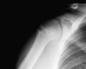
Radiographic visualization of the physis injury is best demonstrated on anteroposterior comparison views of the proximal humerus with the arm internally and externally rotated . External rotation views show widening of the physis at the lateral aspect ( Fig. 1 ). Internal rotational views demonstrate two horizontal radiolucent lines . Lateral fragmentation, calcification, sclerosis, and cyst changes also can be seen, with the widening physis typifying its chronic nature . The exact mechanism of physis injury has yet to be determined; however, it is thought to resemble a Salter Harris type 1 fracture with separation of the metaphysic from the epiphysis. Some investigators have postulated that chronic stress injury leads to an alteration in the endochondral ossification center and delay in apoptosis.
Yamamoto and colleagues recently analyzed humeral retroversion in adolescent baseball players and found a significantly increased amount of retroversion in the dominant arm compared with the nondominant arm. It is generally thought that with normal development the amount of retroversion should decrease with age. Yamamoto and colleagues contributed their findings to a delay in derotation of the humerus caused by repetitive throwing activities. They also found that players who began pitching before age 11 had a greater degree of retroversion (although not statistically significant) than players who started after age 11. These findings, along with radiographic evidence of degenerative changes, point more toward a gradual microtrauma mechanism than an acute fracture. As a result, it is not uncommon for radiographic evidence of closure to take 9 to 12 months, which distinguishes it from a typical (acute) Salter Harris fracture .
Treatment is generally based on professional experience rather than rigorous studies. Eliminating painful activities while allowing the athlete to use other positions or cross-train until pain free is typical. Because radiographic evidence of physis closure can take several months, it is typically not used as a marker of when to return to offending activities. Once patients can perform over-head activities in a pain-free manner, they are gradually allowed to return to play. Physical therapy is usually not needed and, in some case reports, leads to worsening of pain . Overall, the best treatment is to prevent injury in the first place. In a recent review by Caine and colleagues , several essential goals were outlined clearly, including proper education of the athlete, parents, coaches, and other training persons and appropriate training practices, skill development, and periodization.
Rotator cuff injuries
In adolescent athletes, injury to the rotator cuff muscles is rare, occurring in less than 1% of athletes younger than 20 years of age . They are usually the result of an acute traumatic event, secondary impingement because of poor muscular/proprioceptive control, or internal impingement caused by tightness of the posterior capsule/shoulder soft tissue structures. Traumatic events typically involve falling onto an outstretched arm or forcibly impacting an immoveable object (ie, hitting the boards in hockey).
Unlike adults, secondary impingement occurs because of nonoutlet diseases leading to external or internal impingement. Nonoutlet diseases include bursal thickening, contracture of the posterior capsule, and instability of the glenohumeral joint . It is believed that subtle instability of the glenohumeral joint and poor muscle control of the rotator cuff muscles cause a deficiency in the compressive forces needed to stabilize the glenohumeral during over-head movements. This instability allows for repeated contact of the supraspinatus and/or subscapularis against the acrominon or coracoacromial arch, respectively. In the case of internal impingement, it is thought to occur as a result of excessive tightness of the posterior shoulder capsule or overlying musculature. Inappropriate biomechanics lead to the inappropriate contact of the posterior region of the supraspinatus and superior aspect of the infraspinatus tendon with the posterior glenoid rim, which results in a partial-thickness tearing of the undersurface (articular side) of these tendons and fraying of the posterior superior glenoid labrum. Athletes with this form of impingement describe posterior shoulder pain along with decreased throwing/hitting endurance, accuracy, and velocity.
Evaluation of adolescent shoulder injuries should include a detailed history and examination of the shoulder complex. In the throwing athlete, care should be taken to review the pitch count, types of pitches, and amount of pitching throughout the year because these factors have been shown to increase the risk of shoulder injury . Physical examination should include inspection, palpation, range-of-motion testing, neurologic assessment, and stress testing of the rotator cuff muscle group, biceps, labrum, and capsule. It is also important to review the biomechanical motions of the shoulder relevant to the specific sport. Itoi and colleagues found that in most patients with rotator cuff tears, location of pain is a poor predictor of which rotator cuff muscle was injured. They found manual muscle testing of the supraspinatus, infraspinatus, and subscapularis to be a more reliable indicator. Motor examination scores of less than 5/5, 4+/5, and 3/5, respectively, correlated well with arthroscopic findings. Similar to adults, imaging of the shoulder complex with MRI or even ultrasound typically identifies the type of tear.
Treatment of rotator cuff pathology depends on the severity of the tear. In general, adolescent athletes can be treated conservatively with cessation of the aggravating injury and a focused therapy program. In pitchers, time should be taken to review the pitch count, types of pitches, and amount of pitching throughout the year. Therapy should focus on strengthening the scapular stabilizers and rotator cuff muscles. The motions should be integrated into a functional analysis of the pitching biomechanics. After any abnormal motions are corrected, athletes may slowly increase their velocity and return to sport as tolerated.
Related posts:
 Strength Training Recommendations for the Young Athlete
Exercise for Preventing Childhood Obesity
Low Back Pain in the Adolescent Athlete
Acute Knee Injuries in Skeletally Immature Athletes
Nutritional Requirements of the Child and Teenage Athlete
Psychologic Stress Related to Injury and Impact on Sport Performance
Strength Training Recommendations for the Young Athlete
Exercise for Preventing Childhood Obesity
Low Back Pain in the Adolescent Athlete
Acute Knee Injuries in Skeletally Immature Athletes
Nutritional Requirements of the Child and Teenage Athlete
Psychologic Stress Related to Injury and Impact on Sport Performance
Stay updated, free articles. Join our Telegram channel

Full access? Get Clinical Tree


