Stephen Thompson, Luke Choi, Stephen Brockmeier, Mark D. Miller
Shoulder and Arm
Regional Anatomy and Surgical Intervals
Regional Anatomy
Osteology
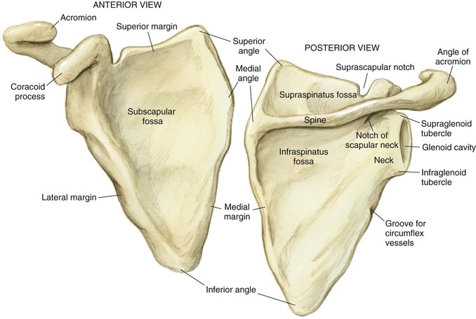
Scapula (Fig. 2-1)
A broad flat bone that serves as an attachment for 17 muscles and 4 ligaments
The scapular spine is the superior aspect of the scapula
The coracoid is the anterior projection that serves as the origin for several muscles and ligaments
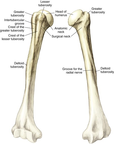
Humerus (Fig. 2-3)
The largest diaphyseal bone in the upper extremity
The hemispherical head is retroverted approximately 30 degrees
The anatomic neck is directly below the head
The surgical neck is approximately 2 cm distal to the anatomic neck
The greater tuberosity is the attachment for the supraspinatus, infraspinatus, and teres minor
The lesser tuberosity is the attachment for the subscapularis
Arthrology
Glenohumeral Joint (Fig. 2-4)
This spheroidal (ball and socket) joint is designed for motion over stability
• Labrum—deepens the socket by 50% and provides a barrier against excessive translation
• Negative intraarticular pressure
• Capsule
• Glenohumeral ligaments (superior, middle, and inferior)
• The anterior band of the inferior glenohumeral ligament is the most important
• Resists inferior translation
• Resists superior translation

Muscles
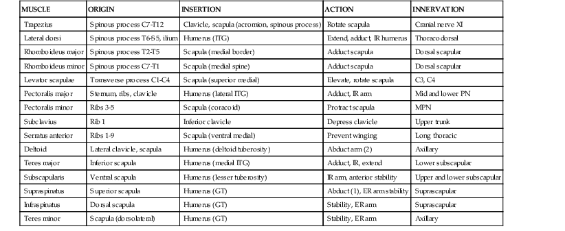
Shoulder Muscle Groups (Fig. 2-6 and Table 2-1)
Connect the upper limb to the axial skeleton
• Trapezius, latissimus, rhomboid major and minor, and levator scapulae
Connect the upper limb to the thoracic wall
• Pectoralis major and minor, subclavius, and serratus anterior
• Deltoid, teres major and minor, supraspinatus, infraspinatus, and subscapularis


Nerves
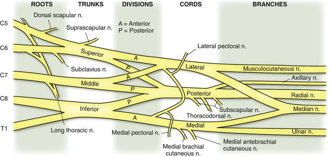
Brachial Plexus (Fig. 2-7)
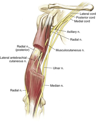
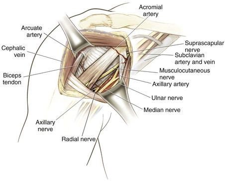
Major Arm Branches (Fig. 2-8)
Musculocutaneous nerve (lateral cord)
• Runs from medial to central anteriorly
• Supplies the biceps (short head), coracobrachialis, and part of the brachialis
• Spirals behind the humerus from medial to lateral
• Supplies the triceps (all three heads) in the arm
Median nerve (medial and lateral cords)
• Runs just medial to the brachial artery in the medial arm
• No major branches in the arm
• Runs just lateral to the brachial artery in the medial arm
Vascularity (Fig. 2-9)
Subclavian Artery
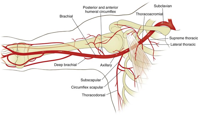
Axillary Artery (Table 2-3)
Three divisions based on the relationship to the pectoralis minor (1, proximal; 2, deep; 3, distal; Table 2-3)
(1) Proximal: supreme thoracic
(2) Deep: thoracoacromial and lateral thoracic (deltoid, acromial, pectoralis, and clavicular)
(3) Distal: subscapular, anterior, and posterior humeral circumflex
Table 2-3
Axillary Artery Branches
| PART | BRANCH | COURSE |
| 1 | Supreme thoracic | Medial to the serratus anterior and pectorals |
| 2 | Thoracoacromial | Four branches (deltoid, acromial, pectoralis, and clavicular) |
| Lateral thoracic | Descends to the serratus anterior | |
| 3 | Subscapular | Two branches (thoracodorsal and circumflex scapular [triangular space]) |
| Anterior humeral circumflex | Blood supply to the humeral head–arcuate artery lateral to the bicipital groove | |
| Posterior humeral circumflex | Branch in the quadrangular space accompanying the axillary nerve |
Cross-Sectional Anatomy
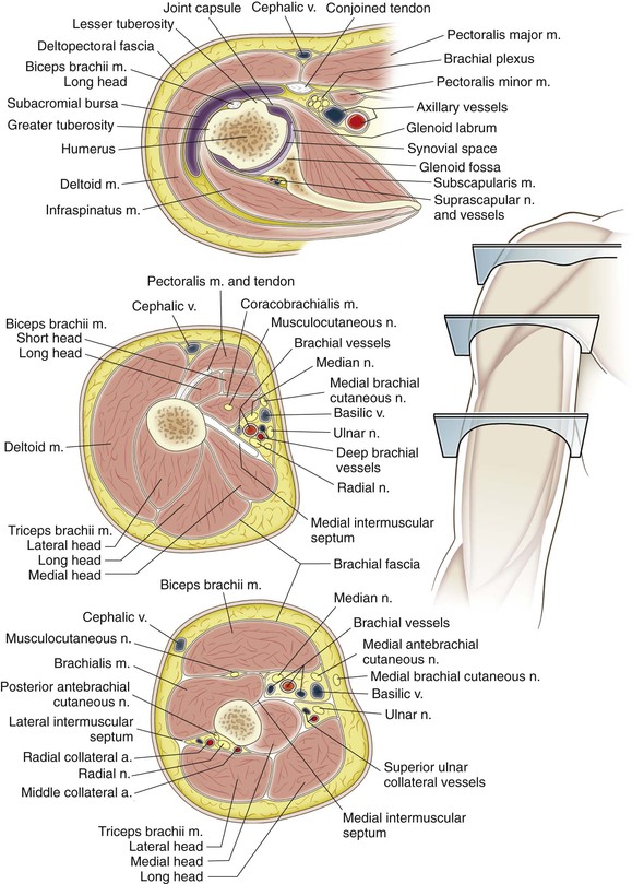
Surgical Intervals
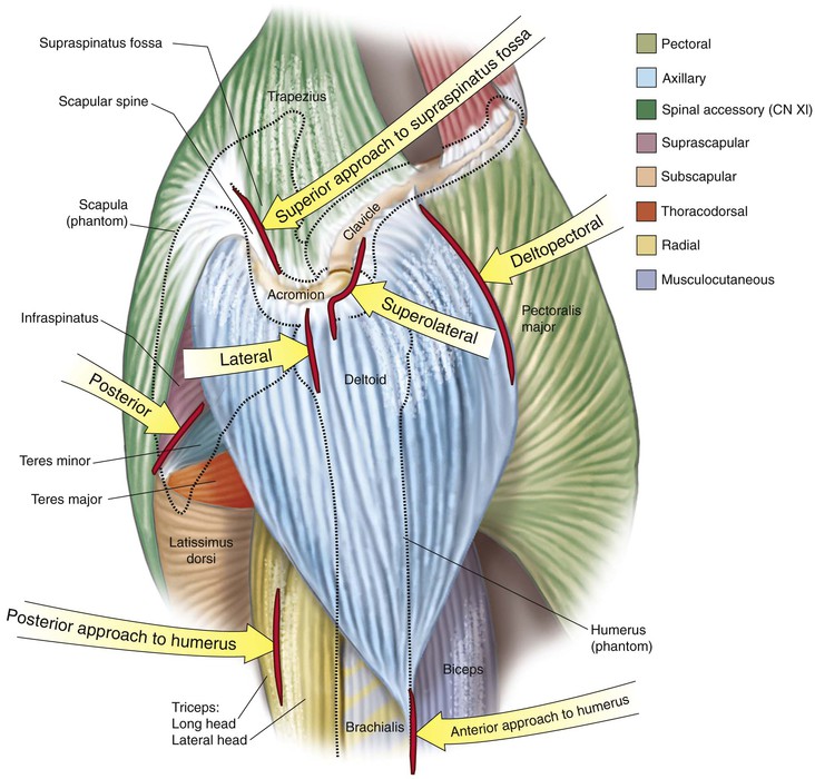
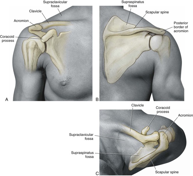
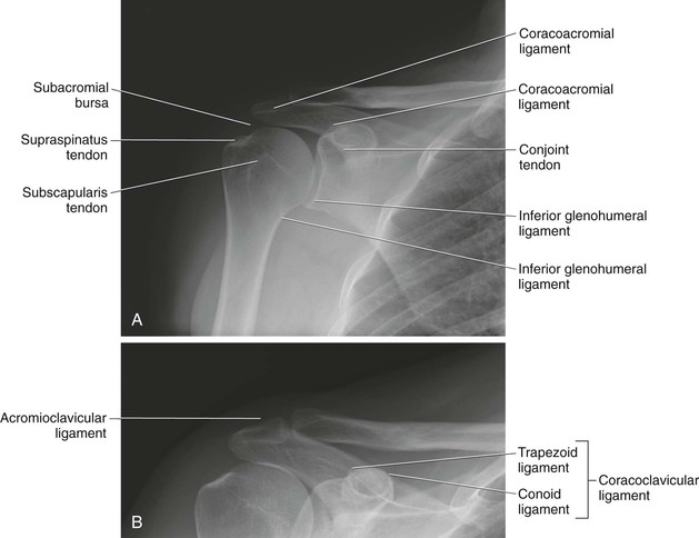
Radiologic Landmarks (Fig. 2-13, A and B)
Hazards
Shoulder (Fig. 2-14)
Nerves
Axillary Nerve
A branch of the posterior cord that supplies the deltoid and teres minor muscles
• Inferiorly as it transverses just below the glenohumeral joint
• Adduct and externally rotate the arm and stay directly on the neck of the glenoid with dissection
• Avoid retractor placement below the subscapularis and capsule
• Palpate the nerve with blunt dissection and use electrocautery without muscle relaxation
• Laterally with any incision or dissection 5 cm or more distal to the lateral acromion
• Place a marking suture at that location, and do not dissect below it
• Posteriorly, in quadrangular space


Stay updated, free articles. Join our Telegram channel

Full access? Get Clinical Tree


