A. Bobby Chhabra, Aaron M. Freilich
Elbow and Forearm
Regional Anatomy and Surgical Intervals
Regional Anatomy
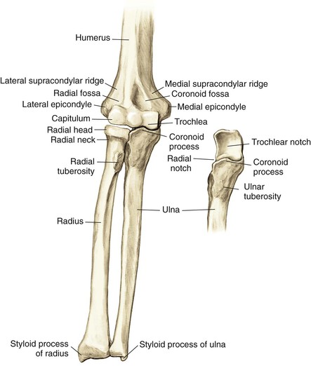
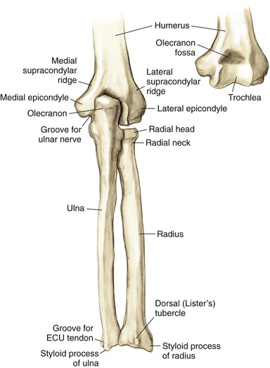
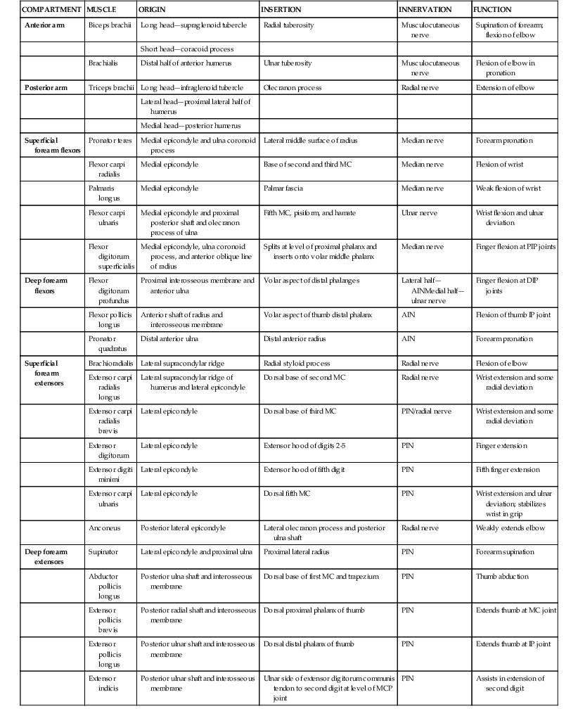
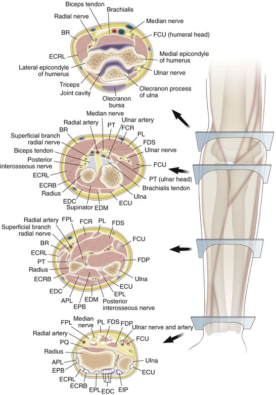
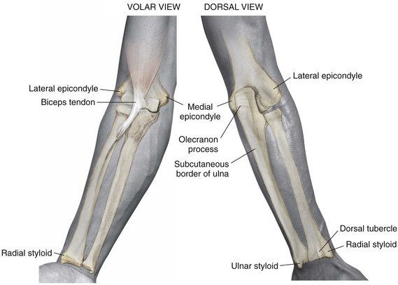
Osteology (Figs. 3-1 and 3-2)
Distal Humerus
Widens and flattens distally into medial and lateral supracondylar ridges, then medial and lateral epicondyles
• The extensor carpi radialis longus (ECRL) originates on the lateral supracondylar ridge
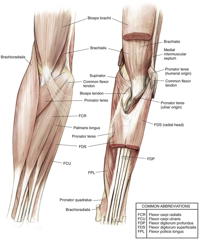
• Common flexor muscles and pronator teres originate on the medial epicondyle
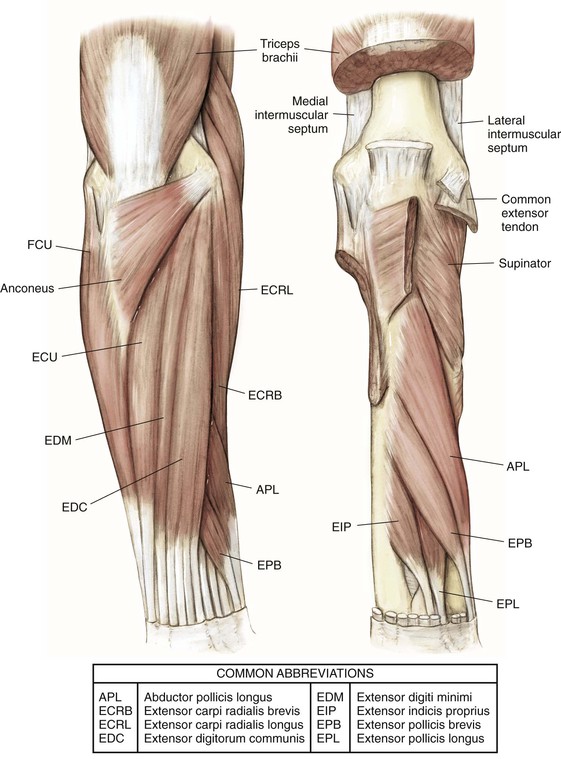
• Common extensor muscles originate on the lateral epicondyle
The capitulum articulates with the radial head laterally
The trochlea articulates with the ulnar trochlear notch medially
The coronoid fossa lies on the anterior humerus
The olecranon fossa lies on the posterior humerus
The radial fossa is anterolateral to accommodate the radial head when the elbow is in flexion
The groove for the ulnar nerve lies between the medial epicondyle and the trochlea
Arthrology (Fig. 3-3)
Elbow Joint
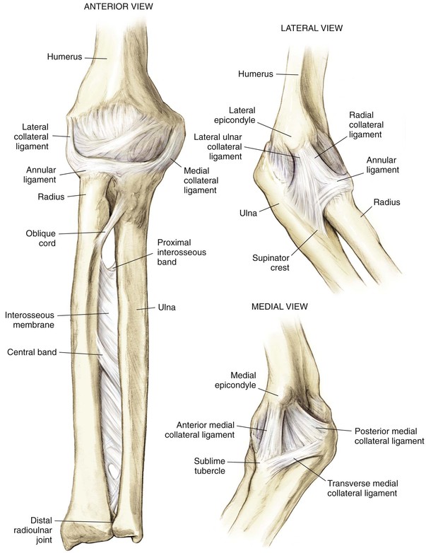
Ligaments
• Annular ligament of the radius
• Lateral ulnar collateral ligament
• From the lateral epicondyle to the ulna supinator crest
• Deficiency leads to posterolateral rotatory instability
• Triangular ligament consisting of three bands
• Anterior band: inferior medial epicondyle to coronoid process
• Posterior band: inferior medial epicondyle to olecranon process
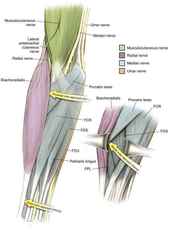
Nerves (Figs. 3-6 and 3-7)
Musculocutaneous Nerve C5, C6, C7
Lies between the biceps brachii and brachialis in the arm
Emerges from beneath the biceps brachii on the lateral side of the biceps tendon
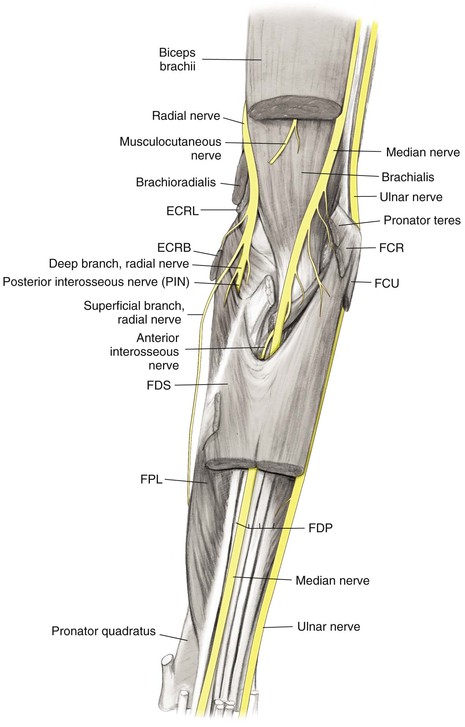
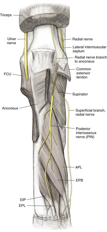
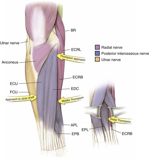
Radial Nerve C5, C6, C7, C8 (T1)
Ulnar Nerve C7, C8 (T1)
Approaches the elbow posteromedially, travels in the medial intermuscular septum of the triceps, and travels beneath the arcade of Struthers
• Crosses the elbow in the cubital tunnel posterior to the medial epicondyle
Travels distally in the forearm between the FCU and the ulnar side of the FDP, supplying both
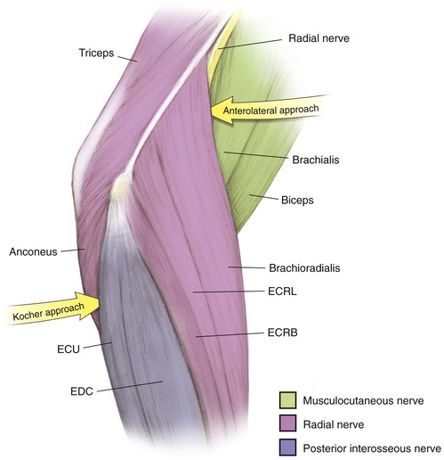
Surgical Intervals
Commonly between internervous planes, allowing for the safest exposure of desired structures; the internervous planes for common approaches to the elbow and forearm are depicted in Figures 3-8 through 3-11

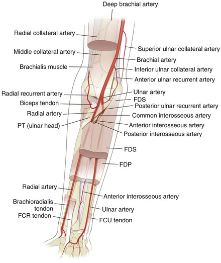
Vascular (Fig. 3-12)
Brachial Artery
The artery branches at approximately the level of the radial neck
• Passes under the bicipital aponeurosis
• Gives off the radial recurrent artery as the first branch
• Gives off the ulnar anterior or posterior recurrent artery, or both, as the first branch
• The common interosseous artery branches at a level just distal to the radial tuberosity, then divides into
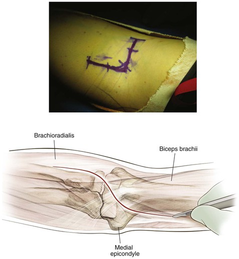
Palpable Anatomic Landmarks for Surgical Incisions and Approaches (Fig. 3-14)
Hazards
Nerves
Radial Nerve
Surgical Approaches to the Elbow
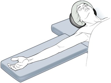
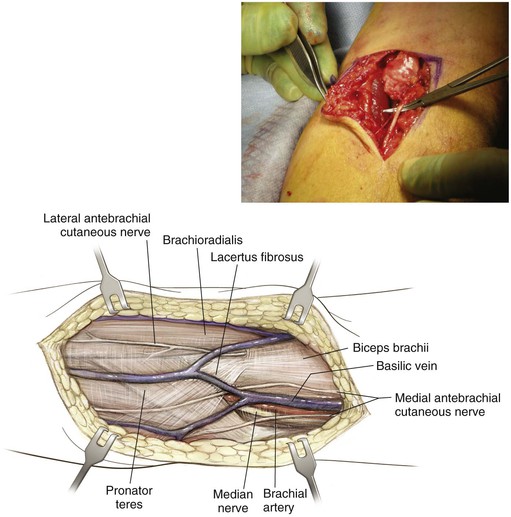
Anterior Approach to the Elbow (Antecubital Fossa)
Indications