Fig. 43.1
Schwannoma of mandible. Panoramic radiograph showing a large lytic lesion at left

Fig. 43.2
Periosteal schwannoma of femur. (a) Radiograph shows a translucent external indentation in the cortical metaphysis at left. (b) CT scan shows a lucent periosteal nodule producing a well-delimited depression in the subjacent cortex
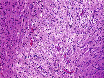
Fig. 43.3
Medium-power microscopic view of a schwannoma with more crowded spindle cell areas (Antoni A) and a looser central area (Antoni B)
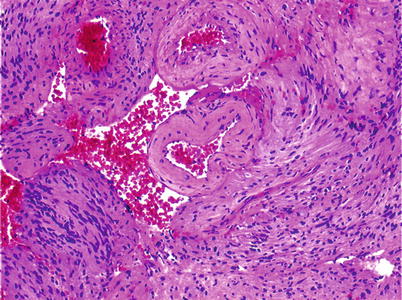
Fig. 43.4


Medium-power microscopic view. Suggestive palisading of the nuclei of the spindle cells, at left, and large congestive blood vessels with thick hyaline walls
Related posts:

Stay updated, free articles. Join our Telegram channel
Full access? Get Clinical Tree
