Fig. 73.1
Rosai-Dorfman disease of the vertebrae. The MRI shows a focal lesion in the vertebral body with extension into the soft tissue
Osseous involvement is sometimes multifocal.
Image Differential Diagnosis
Metastatic carcinoma
Any primary bone neoplasm or infection
Pathology
Histologically, the histiocytes have large vesicular nuclei and abundant foamy cytoplasm.
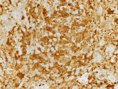
These cells are positive with the S-100 stain (Fig. 73.2).

Fig. 73.2
S-100 stain of the cells of Rosai-Dorfman disease. There is diffuse positivity
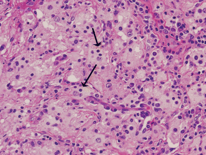
A characteristic feature of the histiocyte in this disorder is emperipolesis, the presence of intracytoplasmic intact lymphocytes (Fig. 73.3). This feature is most prominent in the involved lymph nodes and is less commonly seen in extranodal sites.

Fig. 73.3
High-power photomicrograph of Rosai-Dorfman disease. There is emperipolesis. Large histiocytic cells have intracytoplasmic lymphocytes (arrows)
Stay updated, free articles. Join our Telegram channel

Full access? Get Clinical Tree


