Rehabilitation of the Athlete’s Shoulder

Chapter 2


Rehabilitation of the Athlete’s Shoulder


Clinical and Surgical Pitfalls


• Postural abnormalities can negatively affect kinetic chain function. These should be addressed initially in rehabilitation.


• Core stability, focusing on both local and global muscles, is critical to the success of kinetic chain–focused rehabilitation.


• Inflexibilities such as pectoralis minor tightness and glenohumeral internal rotation deficit need to be assessed and treated.


• The serratus anterior is a multifunctional muscle that is imperative to the stability and mobility of the scapula. Focusing on it solely as a protractor can lead to poor rehabilitation outcomes.


• The lower trapezius is a key muscle in stabilizing the abducted arm.


• Muscle endurance should not be overlooked. High-repetition, low-load exercise will help develop muscle endurance but should be attempted after kinetic chain deficits have been corrected.


Advances in understanding the pathology and appropriate implementation of treatment for shoulder joint injuries have resulted in greater success in defining and restoring the anatomic lesions associated with shoulder dysfunction. Similarly, advances in understanding and implementation of rehabilitation principles for the shoulder have resulted in greater success in restoring the physiologic and biomechanical alterations associated with shoulder dysfunction.


Rehabilitation programs for the shoulder should focus on restoration of functional ability rather than solely on resolution of symptoms. The orthopedic surgeon and the physical therapist must identify and treat all of the structures that are limiting this functional return. Rehabilitation of the shoulder can be both difficult and complex. Multiple joints are involved, and shoulder pain or the pathology that is causing it may actually be the result of distant body biomechanical and physiologic alterations.


This chapter discusses the roles of the orthopedic surgeon in shoulder rehabilitation, presents the basic science in physiology and biomechanics as the basis for shoulder rehabilitation, and offers guidelines and clinical practices that implement the rehabilitation protocols.


The Orthopedic Surgeon’s Roles in Rehabilitation


The orthopedic surgeon plays several roles in shoulder rehabilitation. The first is to understand the basic principles of the kinetic chain. The biomechanical model for striking and throwing sports is an open-ended kinetic chain of segments that work in a proximal-to-distal sequence.1,2 The proximal segment contributions are key components of the sequential activation of body segments that is necessary to accomplish any athletic activity. The kinetic chain harmonizes the interdependent segments to produce a desired result at the distal segment. The goal of the kinetic chain activation sequence is to impart maximum velocity or force through the distal segment (the hand) to the ball, racquet, or other implement. The shoulder does not function in isolation but as a link in the kinetic chain activity that optimizes shoulder function. Alterations in any of the kinetic chain links can affect the shoulder, and alterations in the shoulder can affect the other kinetic chain links. The ultimate velocity of the distal segment is highly dependent on the velocity of the proximal segments. The proximal segments accelerate the entire chain and sequentially transfer force and energy to the next distal segment.25 Because of their large relative mass, the proximal segments are responsible for most of the force and kinetic energy that are generated in the kinetic chain.1 As a result, lower extremity force production is more highly correlated with ball velocity than is upper extremity force production.6


This interaction creates two implications for shoulder rehabilitation. First, the evaluation and identification process preceding shoulder treatment and rehabilitation should include more than just local shoulder structures. The evaluation process should result in a complete and accurate diagnosis of all of the altered structures throughout the kinetic chain. Second, optimum restoration of shoulder function requires that all the kinetic chain segment interactions are reestablished to meet the individual’s needs that existed before injury.


The second role of the orthopedic surgeon is to establish the complete and accurate diagnosis of the anatomic, biomechanical, and physiologic alterations that may be present in shoulder injury and dysfunction. This may seem obvious but is sometimes difficult to implement unless the entire kinetic chain is screened for alterations. Nonetheless, the actual shoulder injury is the primary factor that determines treatment and rehabilitation. This may involve tendon injury or tear, instability, or joint internal derangement, whose overt clinical symptoms can be evaluated by standard diagnostic methods. However, both nonovert local and distant alterations are frequently associated with shoulder clinical symptoms and dysfunction.


The most common local alterations are decreased shoulder internal rotation,7 which creates altered glenohumeral translations,7,8 altered strength or strength balance,7 and alterations in scapular motion and position (scapular dyskinesis).7,911 Inappropriate scapular motion can disrupt the normal smooth coupling of scapulohumeral movement in voluntary activation and is present in most patients with shoulder impingement. Distant alterations include lumbar muscle inflexibility and muscle weakness,12 as well as hip and knee inflexibility.13 Because these alterations are common findings in shoulder injury, they need to be assessed through a screening process in the clinical evaluation. This requires a clinical evaluation approach known as “victims and culprits,” in which the site of symptoms is the “victim,” but the “culprits” may include alterations at other sites.


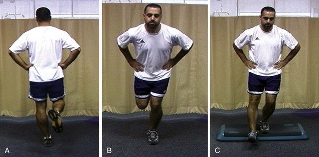
The clinical evaluation should include screening tests for hip and trunk posture and functional strength. Our screening examination includes standing posture evaluation of legs and lumbar, thoracic, and cervical spine; bilateral hip range-of-motion assessment; trunk flexibility assessment; and a one-leg stability series (Fig. 2-1), which assesses control of the trunk over the leg. Any observed abnormalities should be further evaluated in more detail.


Scapular evaluation is performed from behind the patient. It should assess resting position, and the examiner should note any asymmetries with respect to bony landmarks of the inferior angle, medial, and superior border. Dynamic motion screening involves evaluation of the same asymmetries of scapular control with weighted arm abduction and forward flexion, in both ascent and descent.14,15 Observation of any of the three types (“yes” indicates that asymmetry is seen, “no” indicates that asymmetry is not seen) correlates highly with actual biomechanical alteration and can be reliably used as a marker of scapular dyskinesis that needs to be addressed in rehabilitation.16


The third role of the orthopedic surgeon is to determine the timing of entry into and exit from the rehabilitation program. In many cases, entry into rehabilitation should be started before surgery to address deficits of flexibility and strength both locally and at distant portions of the kinetic chain. In many cases, rehabilitation interventions focused on addressing the correct culprits may prevent the need for surgical intervention Postoperative entry into rehabilitation may be quite early, while the shoulder is still protected. Kinetic chain rehabilitation of the legs, trunk, and scapula may be started early, and closed-chain range of motion may be started in safe ranges determined at the time of surgery. Exit from rehabilitation should be based not only on healing of the anatomic lesion, but on normalization of physiology and biomechanics to allow functional return to the demands of the sport or activity. This requires frequent functional assessment as rehabilitation proceeds. Key functional components include range of motion, balanced strength, intact kinetic chain, and functional activities.


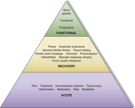
The fourth role is to be familiar with the phases of the rehabilitation program, the content and goals of each stage, and the criteria for progression between stages. Rehabilitation may be divided into three phases (acute, recovery, and functional) based on anatomic injury, anatomic healing, functional capabilities, and functional tasks (Fig. 2-2).


Stay updated, free articles. Join our Telegram channel

Sep 11, 2016 | Posted by in SPORT MEDICINE | Comments Off on Rehabilitation of the Athlete’s Shoulder

Full access? Get Clinical Tree

Get Clinical Tree app for offline access