FIGURE 6.43 Right hand anatomy. (From Tank PW, Gest TR. Lippincott Williams & Wilkins Atlas of Anatomy. Philadelphia, PA: Lippincott Williams & Wilkins, 2009.)
PATIENT POSITION
- Supine on the examination table with the head of the bed elevated 30 degrees.
- The affected wrist is held in a neutral position. The thumb is directed superiorly midway between supination and pronation.
- The wrist is supported with the placement of chucks pads or towels.
- Rotate the patient’s head away from the side that is being injected. This minimizes anxiety and pain perception.
LANDMARKS
1. With the patient supine on the examination table, the clinician stands lateral to the affected wrist.
2. Identify tenderness located in the tendon sheath that contains the abductor pollicis longus and the extensor pollicis brevis.
3. The injection point is located directly between these two tendons. Mark this spot.
4. At that site, press firmly on the skin with the retracted tip of a ballpoint pen. This indention represents the entry point for the needle.
5. After the landmarks are identified, the patient should not move the wrist or thumb.
ANESTHESIA
- Local anesthesia of the skin using topical vapocoolant spray.
EQUIPMENT
- 3-mL syringe
- 25-gauge, 5/8-in. needle
- 0.5 mL of 1% lidocaine without epinephrine
- 0.25 mL of the steroid solution (10 mg of triamcinolone acetonide)
- One alcohol prep pad
- Two povidone–iodine prep pads
- Sterile gauze pads
- Sterile adhesive bandage
- Nonsterile, clean chucks pad
TECHNIQUE
1. Prep the insertion site with alcohol followed by the povidone–iodine pads.
2. Achieve good local anesthesia by using topical vapocoolant spray.
3. Position the needle and syringe at a 45-degree angle to the skin with the needle tip directed proximally.
4. Using the no-touch technique, introduce the needle at the insertion site (Fig. 6.44).
5. Advance the needle toward the convergence of the abductor pollicis longus and the extensor pollicis brevis tendons until the needle tip is located between the tendons in the tendon sheath.
6. Slowly inject the steroid solution as a bolus into the tendon sheath. A small bulge in the shape of a sausage should develop in the tendon sheath.
7. Following injection of the corticosteroid solution, withdraw the needle.
8. Apply a sterile adhesive bandage.
9. Instruct the patient to move his or her thumb through its full range of motion. This movement distributes the steroid solution throughout the tenosynovial sheath.
10. Reexamine the hand and wrist in 5 min to confirm pain relief.
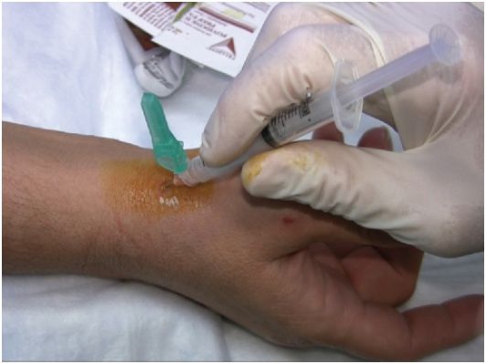
FIGURE 6.44 de Quervain’s tenosynovitis injection.


