Proximal Femoral Osteotomy in the Skeletally Immature Patient With Deformity

CHAPTER 36 Proximal Femoral Osteotomy in the Skeletally Immature Patient With Deformity


Basic science


A varus-producing proximal femoral varus osteotomy (PFO) that creates a neck shaft angle of 100 degrees to 110 degrees is indicated to correct coxa valga for the treatment of hip dysplasia in the young child. The secondary remodeling of the neck shaft angle to near normal (i.e., 130 degrees) is anticipated, as is the overgrowth of the femur, which results in a limb-length inequality (i.e., long on the operated side).


When correcting valgus deformities with a varus-producing osteotomy, the osteotomy and the point of correction are typically distal to the site of deformity (i.e., the head–neck junction). Consequently, undesirable medial deviation of the mechanical axis of the lower extremity can occur. To minimize this occurrence, the distal fragment may be displaced medially to maintain the passage of the lower-extremity mechanical axis through the center of the knee joint. Similarly, when performing a valgus-producing PFO to correct coxa vara, fixation that displaces the distal fragment medially may result in the lateral deviation of the lower-extremity mechanical axis, which potentiates a genu valgum deformity. This can be prevented by ensuring that the distal fragment is translated and fixed lateral to the proximal fragment. When correcting varus or valgus deformities in older children or adolescents with the use of a PFO, the distal fragment should be aligned with the piriformis fossa; this will facilitate the passage of the stem of the femoral prosthesis if a total hip replacement becomes necessary later in life. This is particularly important when a PFO is used to correct a severe deformity that results from a chronic slipped capital femoral epiphysis (SCFE) in which the correction is intertrochanteric and distal to the site of the deformity in the physis.


Valgus-producing osteotomies typically lengthen the lower extremity, whereas varus osteotomies shorten the extremity. Abductor length also changes in relation to the position of the greater trochanter. When correcting varus deformities with a proximal valgus osteotomy, the tip of the trochanter—which is cartilaginous in younger patients and bony in older patients—should be at the level of the center of the femoral head. This may not be possible if the femoral neck is very short. In older children, the distal transfer of the greater trochanter can be performed to accomplish this. Similarly, when performing a varus osteotomy, the resulting location of the tip of the trochanter varies with the underlying pathology and the amount of varus introduced. The tip of the greater trochanter may be normally located so that it is level with the center of the head after the correction of coxa valga or proximal to the center of the head after PFO for containment treatment of Legg-Calvé-Perthes disease. In the former situation, the tip of the trochanter was below the center of the head because of an increased neck shaft angle in a valgus deformity; in the latter situation, the neck shaft angle was normal before the osteotomy, so the varus PFO raises the level of the greater trochanter.


Nonunion after osteotomy of the proximal femur in children is very unlikely. Those with underlying neuromuscular disorders (e.g., static encephalopathy, myelodysplasia) may have delayed healing, which can lead to a loss of correction. Among older children (i.e., those who are more than 10 years old), adequate rigid fixation is essential to ensure healing without a loss of correction. However, there are limitations to the stability of a blade plate and osteotomy construct that may compromise healing. This is more likely after an osteotomy that is performed to correct a very severe deformity, such as SCFE in a large adolescent, in which case it may be difficult to establish adequate contact of the osteotomy fragments and provide sufficient deformity correction. Morbid obesity (i.e., a body mass index of more than 40) is a frequent risk factor for patients with severe SCFE, particularly in North America. Deformity may persist at any age in a child as a result of undercorrection. Deformity may also recur because of unpredictable growth patterns, particularly among young children.


Knowledge of the vascular supply to the proximal femur is important to avoid avascular necrosis as a complication. The medial femoral circumflex artery takes its origin from the deep femoral artery. It passes just medial to the proximal femur at the level of the iliopsoas tendon and then courses posteriorly to its entry into the proximal posterior lateral femoral neck. Injury to this vessel is possible when performing an intertrochanteric osteotomy if there is excessive medial penetration with either a saw or osteotome. Similarly, the terminal branches of the medial circumflex vessel can be injured at the base of the femoral neck while performing a femoral neck osteotomy or an osteotomy of the greater trochanter.


Brief history and physical examination


The history and physical examination findings of patients who are candidates for a PFO vary with the etiology of the underlying hip pathology. A growing child with hip dysplasia usually has no complaints and presents with minimal if any abnormalities during the examination. A limp is often not noted, and the range of motion may be normal. The decision to perform a PFO is based solely on persistent abnormal radiographic findings. The history of prior treatment for DDH, however, may be important to explain the proximal femoral deformity that occurs as a result of avascular necrosis. Limb-length inequality should be taken into consideration when performing a PFO (i.e., varus shortens, valgus lengthens); this may influence the need for temporary shoe lifts or later limb-length equalization. Patients with coxa vara often limp because of limb shortening or abductor weakness, which produces a Trendelenburg gait. These patients tend to walk with an external rotation deformity. Children with Legg-Calvé-Perthes disease have a history of intermittent limp and pain that may be anterior hip pain or referred pain in the distal thigh. The adolescent patient who presents with a proximal femoral deformity as a result of an SCFE will have a history of noted functional hip joint disability as well as intermittent pain that is often in the distal thigh and reported as knee pain. These patients walk with an externally rotated lower-extremity deformity and often sit with limited hip flexion and abduction. When symptomatic, these patients present for evaluation complaining of hip pain, particularly with activities that require flexibility. Adolescent patients who are being considered for a PFO may present with morbid obesity. Anesthetic consultation may be indicated for patients with a history of sleep apnea, a body mass index of more than 40, or both. The families of children with excessive version express concern about the appearance of their gait as well as their clumsiness.


Imaging and diagnostics


Appropriate imaging of the pelvis, hip joint, and proximal femur is essential during preoperative planning to ensure a satisfactory outcome after osteotomy treatment of hip joint pathology. The initial radiographic evaluation should include both standing anteroposterior and supine frog-leg lateral views. Any pelvic tilt and associated limb-length discrepancy (LLD) should be noted. When planning for the osteotomy, it may be helpful to repeat the standing pelvic x-ray and to note the lift necessary to balance the pelvis. Functional radiographs with the hip in variable combinations of flexion and extension, abduction and adduction, and internal and external rotation can be helpful when determining the best position for optimal hip joint congruity and developing the strategy for the performance of the osteotomy. The potential effect of a varus osteotomy to redirect the femoral head into the acetabulum can be assessed with the use of a Von Rosen view (i.e., a supine anteroposterior view with the hips in flexion, abduction, and internal rotation). By contrast, a supine x-ray with the hip adducted can demonstrate the potential effect of a proximal femoral valgus osteotomy. Sagittal plane deformities—such as those seen with DDH, Legg-Calvé-Perthes disease, and coxa valga—can readily be seen on the frog-leg lateral views. To better identify the pathology with an severe deformity such as the posterior tilt of the epiphysis seen with an SCFE, a cross-table lateral view with the hip in 15 degrees of internal rotation should also be obtained. On occasion, it may be helpful to obtain a computed tomography scan for a more detailed image of the pathologic anatomy. The use of three-dimensional reconstruction is helpful for addressing complex femoroacetabular deformities. Whether a three-dimensional computed tomography scan would provide a better understanding of the bony deformity that could optimize the outcome of surgical treatment must be weighed against the increased radiation exposure for the patient. Alternatively, magnetic resonance imaging can be used to further define the proximal femoral and hip joint morphology and to facilitate the planning of the optimal surgical strategy.


Surgical technique


Although the indications and ages of patients who are candidates for PFO are quite variable, the surgical anatomy and the basic technique for performing a PFO are quite similar. In both young and older children, PFOs are typically performed with the patient in the supine position on a radiolucent table. The patient is positioned so that the involved hip and thigh are close to the edge of the table and the operating surgeon. A soft bump (lift) is placed under the operative hip. For the smaller child, a rolled sheet is positioned against the opposite side of the patient to minimize the patient’s sliding away from the surgeon during the procedure. The ipsilateral lower torso and leg are prepped down to the ankle. A stockinet covers the extremity from the toes to the mid thigh. Drapes are secured and sealed to the skin with an adhesive barrier material. The lateral approach is used for most PFOs.


Surgical Technique for the Lateral Approach to the Proximal Femur


The lateral surgical approach is similar for all age groups. A straight lateral incision that extends from the tip of the trochanter to a point several centimeters distal to the greater trochanter is used for exposure. The length of the incision varies with the size of the implant to be used for fixation and with the size of the child. Sharp dissection is carried down to the fascia lata with the scalpel and electrocautery. The skin and subcutaneous tissue are elevated together off of the fascia lata for a few centimeters in both the anterior and posterior directions. The fascia lata is divided in line with the skin incision. Just deep to the fascia lata lies the fascia of the vastus lateralis, which is longitudinally incised directly over the vastus lateralis muscle.


With lateral traction on the posterior edge of the vastus lateralis fascia and counter (medial) traction on the vastus lateralis muscle mass, the electrocautery is used to mobilize and reflect the vastus muscle from lateral to medial off of the lateral intermuscular septum. Care is taken to avoid dissecting through the septum and into the posterior muscle compartment of the thigh. Proximally, the vastus release extends anteriorly over the shaft of the femur. The lateral-to-medial reflection of the vastus lateralis muscle mass is completed from proximal to distal. Within the distal vastus muscle, care should be taken to identify and coagulate the vessels that distally perforate through the lateral intermuscular septum from the posterior to the anterior compartment.


As the vastus lateralis is retracted medially, the periosteum of the femur is visualized. The lateral periosteum is incised, and the femur is subperiosteally exposed just distal to the lesser trochanter. A curved retractor is carefully placed subperiosteally around the anterior shaft of the femur. Posteriorly, the periosteum and the soft tissues are more securely attached to the bone; they are best elevated off of the bone with the electrocautery and the elevator. With blunt dissection, the subperiosteal exposure can be extended distally. With an adequate exposure, the femur should be visible and accessible from the proximal aspect of the greater trochanter to a point distal to the lesser trochanter, which is sufficient for the application of the fixation plate (Box 36-1).


Surgical Technique for Correcting Proximal Femoral Valgus Deformity in Young Children


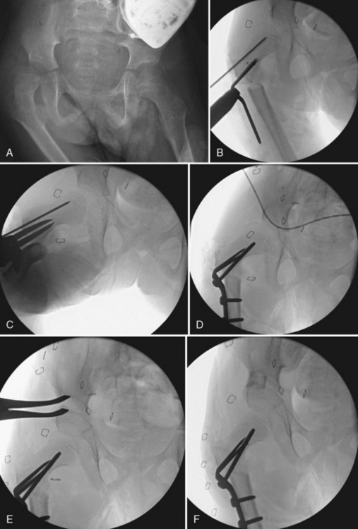
For children who are less than 5 years old, a small fragment, a semi-tubular straight plate, a Wagner forked plate (Aesculap, San Francisco, CA), or a small blade plate (Synthes, Paoli, PA) can be used for fixation. The appropriate implant is temporarily inserted into the incision to assess the adequacy of the exposure. If necessary, the skin and fascial incisions are extended distally to facilitate the insertion of the plate. C-arm imaging is used to confirm the optimum position of hip abduction, flexion, and internal rotation needed to reduce or satisfactorily position the femoral head into the acetabulum. A four- or five-hole small-fragment straight semi-tubular plate is the best choice for a femoral shortening osteotomy in conjunction with the open reduction of a developmentally dislocated hip. The anterior iliofemoral approach and open reduction are performed first through an oblique incision that parallels the lateral edge of the iliac crest. For the shortening osteotomy, the lateral proximal femur is approached as previously described. The osteotomy (Figure 36-1) is performed just distal to the lesser trochanter. The plate is placed just distal to the inferior edge of the greater trochanter physis as assessed with the C-arm. The plate is provisionally fixed by inserting a screw in the most proximal screw hole. A drill hole is also made in the bone that corresponds with the second most proximal hole. The proximal screw is loosened, and the plate is rotated anteriorly. The femoral osteotomy is performed just distal to the predrilled second proximal hole.


With the femoral head reduced, the amount of fragment overlap is directly measured; it is typically 1 cm to 2 cm. This determines how much shortening is required. A second osteotomy that is parallel to the first is completed. The fragments are reduced and fixed to each other with the correction of excess anteversion as appropriate. The femoral head is reduced, and the position and stability are assessed. If additional shortening or a change in the rotational alignment is needed, the distal screws are removed, the distal fragment is further shortened or rotated, and the plate is reapplied with the use of new holes.


A Wagner forked plate works well for younger, smaller patients as fixation if angular correction is desired beyond shortening or rotation, as in a varus osteotomy to correct coxa valga associated with residual DDH or with static encephalopathy. If the Wagner plate is to be used, a smooth K-wire is inserted just proximal to the intended site of plate insertion (Figure 36-2). The K-wire is inserted from lateral to medial and placed superiorly in the neck on the anteroposterior view to provide enough space for the subsequent insertion of the Wagner plate. It is centered in the femoral neck on the lateral view. Subperiosteal retractors are circumferentially placed around the femur to protect the soft tissues. A femoral osteotomy is performed at the midpoint of the lesser trochanter, and this is confirmed with the use of C-arm imaging. The femur is transversely cut with an oscillating power saw under direct vision. After completing the osteotomy, consideration should be given to shortening the distal fragment 1 cm to 2 cm, because growth stimulation and secondary limb-length discrepancy often occur in these young patients. A Wagner forked plate of appropriate size is inserted into the proximal femoral fragment with the use of the previously inserted K-wire as a guide. The forked tips of the plate are inserted through the distal lateral cortex and carefully advanced medially and proximally into the femoral neck; the correct position is confirmed with the C-arm. After the prongs of the plate are fully inserted, the distal femoral fragment is reduced and secured to the proximal fragment with a self-centering bone-holding clamp. When performing a varus-producing osteotomy, the distal fragment should be slightly medialized relative to the proximal fragment. The proportion of medialization will vary slightly, depending on the entry point of the plate in the proximal fragment. Excessive medialization can produce an unstable construct as a result of a loss of contact between the fragments.


The reduction of the femoral head in the acetabulum and the neck shaft angle is assessed with the use of C-arm imaging. If necessary, the varus/valgus alignment can be adjusted by removing the bone-reduction clamp and bending the side plate in situ. In small children (i.e., those less than 3 years old) a varus neck shaft angle of 105 degrees to 110 degrees will typically remodel into an acceptable neck shaft angle at maturity. Obtaining an excessive neck shaft angle (i.e., more than 90 degrees) is undesirable. Remodeling occurs slowly, and a near-normal neck shaft angle may never be achieved. A neck shaft angle of 110 degrees to 115 degrees is used in older children, who have less remodeling potential. The rotational alignment of the two fragments is adjusted to help optimize the reduction of the femoral head into the acetabulum. By doing so, excess external rotation of the distal fragment should be avoided, because this may lead to an out-toeing gait. It may be desirable to slightly extend the distal fragment to create flexion of the proximal fragment, which provides anterior femoral head coverage by the acetabulum in the weight-bearing position. The extension of the distal fragment is achieved by tilting the plate posteriorly as the prongs are inserted into the proximal fragment.


The hip range of motion should be assessed, with flexion and extension, abduction and adduction, and internal and external rotation being noted; passive abduction should be at least 30 degrees. If passive hip abduction motion is limited by an adductor muscle contracture, which is likely with neuromuscular hip dysplasia, an adductor tenotomy may be indicated; this is typically performed at the beginning of the surgical procedure. Hip rotation motion should be assessed, and external and internal rotation should be approximately equal. After a satisfactory reduction of the fragments is achieved, the plate is secured to the distal femur with two bicortical screws. The most proximal hole on the plate should be filled with one long screw directed into the femoral neck to securely engage the proximal fragment.


The final neck shaft angle, the plate fixation, and the hip reduction should be evaluated using the C-arm. A deep suction drain is placed, and the wound is closed in separate fascial layers. For young children (i.e., those less than 6 years old), a 1½ hip spica cast holding the thigh in a slightly abducted and flexed position is applied for 5 to 6 weeks (Box 36-2).


Stay updated, free articles. Join our Telegram channel

Jul 24, 2016 | Posted by in MUSCULOSKELETAL MEDICINE | Comments Off on Proximal Femoral Osteotomy in the Skeletally Immature Patient With Deformity

Full access? Get Clinical Tree

Get Clinical Tree app for offline access