Prolonged Drainage, Skin Necrosis, and Wound Problems


Prolonged Drainage, Skin Necrosis, and Wound Problems


Michael D. Ries


INTRODUCTION

Serous wound drainage after total knee arthroplasty (TKA) is not uncommon, but prolonged drainage is associated with an increased risk of developing infection. If infection is suspected, early irrigation and debridement is indicated to prevent chronic infection involving the prosthetic components.

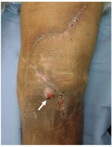
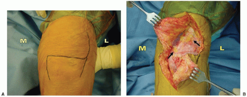
Skin necrosis can rapidly lead to exposure of the implant and deep infection of the TKA. The risk of skin necrosis can be reduced by utilizing previous scars and more lateral incisions in the multiply scarred knee when possible, and maintaining full-thickness skin and subcutaneous flaps during surgical exposure, as well as limiting early knee range of motion (ROM) after surgery. If full-thickness necrosis occurs, with resultant exposure of the TKA, prompt debridement and soft tissue coverage is necessary. Most soft tissue defects can be covered successfully with a medial gastrocnemius muscle flap transposition.


POSTOPERATIVE WOUND DRAINAGE

Blood drainage from the wound after TKA is common during the first few days after surgery. Once the wound is sealed and the skin begins to epithelialize, drainage should diminish considerably. However, serous fluid drainage can continue for up to a week in some patients. The fluid may originate from edema, blood debris, and ischemia in the subcutaneous tissues. When drainage is present, the wound is at a higher risk of developing infection (1,2). Infection is more likely to occur in patients with poor vascularity of the soft tissues, diabetes, immunosuppressive disorders, malnutrition, steroid use, and in obese patients with large subcutaneous tissue planes exposed during surgery (3,4).

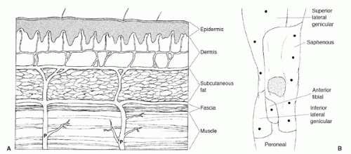
Surgical technique can affect the vascularity to the skin and subcutaneous layer. Ideally, the skin incision is placed in close proximity to the arthrotomy incision to avoid elevating soft tissue flaps. However, prior scars often restrict the location of the surgical incision so that flaps are required to expose the medial retinaculum and permit use of a medial parapatellar arthrotomy. Blood vessels perforate through the fascia to form an anastomosis deep to the subcutaneous layer, which supplies circulation to the subcutaneous tissue and skin (Fig. 33-1) (5). Dissection of the subcutaneous tissue should be performed along the subfascial plane between the deepest portion of the subcutaneous layer and deep fascia. If full-thickness skin and subcutaneous soft tissue flaps are dissected from the fascial layer, relatively large flaps can be raised without compromising soft tissue vascularity. If dissection is carried out into the subcutaneous plane and subcutaneous tissue is left attached to the fascia, however, the overlying soft tissue can become devascularized and lead to ischemia or necrosis (Fig. 33-2).


FIGURE 33-1 (A) The microvascular circulation to the skin and subcutaneous tissue consists of deep perforating vessels (P) that penetrate the fascia and form an anastomosis along the deep portion of the subcutaneous layer. Skin flaps should be raised by dissection along the fascial plane to maintain the anastomotic blood supply to the subcutaneous tissue and skin. (B) The areas of the skin over the knee supplied by the deep perforators are shown. Most of the blood supply comes from the medial side, so the lateral skin flap will be more hypoxic than the medial skin flap, and using a more medial incision may increase the risk of skin necrosis. (Reprinted with permission from Younger ASE, Duncan CP, Masri BA. Surgical exposures in revision total knee arthroplasty. J Am Acad Orthop Surg 1998;6:55-64.)


FIGURE 33-2 A 54-year-old man underwent total knee arthroplasty for posttraumatic arthritis. Three weeks after surgery, a small amount of drainage developed lateral to the surgical incision. Surgical exploration demonstrated deep infection.


TABLE 33-1. Summary of Types of Acute Postoperative Wound Drainage


Diagnosis


Source of Drainage


Signs


Infected (yes/no)


Treatment


Type 1 Superficial serous drainage


Edema caused by surgical trauma and ischemia of subcutaneous soft tissues.


After initial onset, serous drainage volume decreases daily.
No cellulitis, or wound induration.


No


Daily sterile dressing changes. Oral or IV antibiotics if drainage excessive to prevent wound colonization.


Type 2
Superficial infection


Abscess fluid in subcutaneous layer.


Serous drainage, volume increases daily.
Cellulitis around wound. Fever, erythema, and induration may be present.


Yes


Joint aspiration to confirm absence of deep infection. If drainage is minimal, treat with IV antibiotics and dressing changes. If drainage persists, then I and D of subcutaneous layer.


Type 3 Deep serous drainage


Intra-articular blood and joint fluid expressed into subcutaneous layer through a fascial defect.


Serous drainage, increases with activity. No fever, erythema, or induration.


No


I and D of joint, place drains, repair fascial defect if possible, use postoperative antibiotics.


Type 4 Deep postoperative infection


Infected hematoma or abscess fluid expressed into subcutaneous layer through a fascial defect.


Thick serous or purulent drainage. Fever, erythema, and induration may be present.


Yes


I and D with tibial insert exchange. Closure of fascial defect over drains. Six weeks post-operative IV antibiotic therapy.


I and D, irrigation and debridement; IV, intravenous.


Wound drainage management depends on the location of fluid accumulation (superficial or deep) and presence or absence of infection (Table 33-1). Serous drainage originating from edema in the subcutaneous layer does not necessarily represent infection, but the expression of fluid through the surgical incision indicates that the wound is not sealed. Incomplete wound healing indicates that the wound does not provide a complete barrier to contamination from the skin. Sterile dressings should be maintained on the wound and changed daily as often as necessary to collect wound drainage.

When drainage is present, the wound is at risk of developing infection from skin contamination (1,2). If the drainage volume decreases daily and clinical signs of infection (fever, erythema, and induration) are absent, however, then the drainage can be treated with dressing changes alone and without antibiotics or surgical debridement. Antibiotics can be used to manage a noninfected draining surgical wound as a prophylactic measure to prevent infection because the wound is incompletely sealed and exposed to skin contamination. However, antibiotic therapy alone for treatment of a deep post- operative infection is not an appropriate method to eradicate infection. If there is a clinical suspicion that deep infection is present and the source of wound drainage is not clear, then use of antibiotics may suppress an infection and make the diagnosis more difficult to establish. Therefore, the distinction between a wound that is not infected and draining from the subcutaneous tissues and one that is infected or draining from the knee joint into the subcutaneous layer is important.


IRRIGATION AND DEBRIDEMENT FOR PROLONGED DRAINAGE


Indications

Irrigation and debridement (I and D) is indicated for deep wound drainage originating within the knee draining out of the skin incision (type 3 or 4, Table 33-1). Drainage from a sinus outside of the surgical incision indicates that drainage is occurring from within the knee joint and surgical debridement is necessary (Fig. 33-3). I and D is also appropriate for superficial infection (type 2, Table 33-1). I and D is not necessary to treat serous fluid drainage resulting from edema or ischemia of the subcutaneous tissue that is not infected (type 1, Table 33-1). However, type 1 drainage may
convert to type 2 drainage. A period of observation is appropriate for type 1 drainage. If the drainage is decreasing each day and if clinical signs of infection are not present, then the drainage should resolve as wound healing occurs in the subcutaneous layer.


FIGURE 33-3 (A) A previous S-shaped scar was present in a patient who required total knee arthroplasty. The medial (M) and lateral (L) sides of the knee are indicated. (B) The previous incision was used, which required elevation of skin and subcutaneous soft tissue flaps from the fascia. Subcutaneous tissue has been left attached to the fascia and detached from the skin flap (arrows) indicating that dissection extended into rather than deep to the subcutaneous layer. As a result, a portion of the overlying skin and subcutaneous soft tissue may become ischemic.


Contraindications

There are virtually no contraindications to I and D for wound drainage suspected to originate from within the knee (type 3 or 4, Table 33-1) or associated with a superficial infection (type 2, Table 33-1). If infection is suspected, I and D is the most appropriate treatment. If I and D is performed during the early postoperative period, however, primary wound closure may be associated with excess skin tension caused by edema and contracture of the soft tissues over the knee. If the wound is closed under tension, limiting early knee ROM after surgery can be beneficial to allow wound healing to occur (6). If primary wound closure is not possible after I and D, additional soft tissue flap coverage is necessary. The integrity of the soft tissues and any potential difficulty with primary wound closure should be carefully assessed prior to I and D.


Technique

If I and D is performed for treatment of superficial infection (type 2, Table 33-1), the fascial layer will be intact, and debridement is limited to the skin and subcutaneous layer. Fluid and soft tissue specimens should be obtained for culture. After debridement, the wound should be closed over a subcutaneous drain.

For debridement of deep wound drainage (type 3 or 4, Table 33-1), the wound should be adequately exposed during I and D to provide access to all areas of the knee including the posterior compartment. The surgeon can perform debridement as an open procedure or arthroscopically. Arthrotomy provides wide exposure of the knee, permits exchange of the tibial insert, and direct exposure of the metallic components to determine whether they are well fixed. Tibial insert exchange allows exposure and debridement of the tibial base plate surface and better exposure of the posterior compartment of the knee. Successful results have also been reported with arthroscopic debridement (7

Only gold members can continue reading. Log In or Register to continue

Stay updated, free articles. Join our Telegram channel

Jun 13, 2016 | Posted by in ORTHOPEDIC | Comments Off on Prolonged Drainage, Skin Necrosis, and Wound Problems

Full access? Get Clinical Tree

Get Clinical Tree app for offline access