Chapter 4 Problematic Stress Fractures of the Foot and Ankle
 Pearl
PearlIntroduction
Stress fractures differ from acute fractures in that their course generally is more gradual and their radiographic appearance can be elusive.1 Stress fractures of the foot and ankle are most common in running athletes, especially those who jump. For example, track athletes, ballet dancers, and basketball players have a high incidence of stress injury. Many studies have implicated biomechanical factors, such as leg-length discrepancies, cavus foot deformities, and limb malalignment. Women have a higher incidence of stress fractures, and amenorrhea often is a concomitant finding in female athletes with these injuries.2
Stress Fracture of the Tarsal Navicular
Anatomy and presentation
The tarsal navicular serves as a keystone in the medial longitudinal arch and consequently is subjected to tremendous forces through the foot. Moreover, nutrient arteries arising from both the anterior and posterior tibial arteries create a generous supply of blood to the medial and lateral thirds of the navicular. The result is a poorly vascularized zone in the middle third of the bone.2 Incredible stresses and decreased nutrition make the middle third of the navicular the most common location for stress fracture.
Misdiagnosis of stress fracture of the tarsal navicular generally is the rule rather than the exception. There are several sources of midfoot pain that are more common, including plantar fasciitis, anterior tibial and posterior tibial tendinitis, spring ligament injury, Lisfranc sprain, and degenerative joint disease.3 Therefore unrelenting symptoms in the seemingly normal midfoot merit further diagnostic workup and probably referral.
Towne et al.4 first reported stress fracture of the tarsal navicular in 1970. In this series of two patients, each was a distance runner who had experienced midfoot pain with swelling and failure to respond to conservative therapy. Plain radiographs were negative, and only specialized studies were able to reveal the occult fracture. Subsequent reports have corroborated a history of insidious pain in the midfoot that is relieved by rest and exacerbated by forceful striking of the forefoot and with direct palpation of the navicular.5–7 A common thread in these reports is the normal appearance of plain radiographs. In most cases the diagnosis must be confirmed by computed tomography (CT), magnetic resonance imaging (MRI), or bone scan.
Imaging
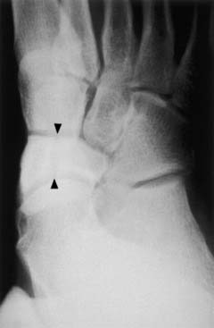
Stress fracture of the tarsal navicular often is overlooked secondary to the low sensitivity of plain radiographs in diagnosing this condition. Some characteristics appreciated on anteroposterior views of the foot have been shown to correlate with navicular stress fractures. These include sclerosis of the proximal border of the navicular; a short first metatarsal; metatarsus adductus and hyperostosis; and stress fracture of the second, third, and fourth metatarsals. Improved imaging techniques have demonstrated that most fractures are linear, lie in the middle third of the navicular, and can be complete or partial.8 Oblique or supinated radiographs can be useful (Fig. 4-1).
Radionuclide bone scanning always demonstrates increased isotope uptake and can be a useful adjunct in the diagnosis of ill-defined midfoot pain. Views should include a medial, lateral, and plantar view. Uptake generally will appear in the shape of the navicular on the plantar view.5 Although radionuclide scanning can assist in localizing the area of concern to the navicular, definitive diagnosis and definition of the fracture pattern require tomography or computer-aided tomography.
The majority of fractures occur in the middle third of the navicular. An anatomic anteroposterior (AP) tomogram views the middle third of the navicular en face and therefore is more likely to identify fractures.5 However, modern computer-aided tomography has supplanted the use of tomography and is essential in the delineation of fracture pattern. Fine, 1.5-mm cuts are necessary in the axial plain to ensure that small incomplete fractures on the dorsal surface of the bone are not “skipped.” The role of MRI has not been clearly defined but likely will prove useful in the early diagnosis of this condition. Successful identification of the injury and its anatomy is crucial to effective management of the fracture.
Treatment
Complete elucidation of the fracture pattern is important in dictating management of the athlete. Patients with incomplete and nondisplaced complete fractures can respond well to conservative management. When treated in a nonweight-bearing cast for at least 6 weeks, 86% to 100% of patients will go on to union.2–4
Patients with displaced fractures or those who have failed nonoperative management benefit from bone grafting with or without open reduction and internal fixation. Most of these athletes will return to sport within 5 to 7 months.2–4 High-performance career athletes and the treating surgeon may elect a more aggressive approach to nondisplaced fractures. Theoretically, surgical management of the injury could expedite the return to sport.
Stress Fracture of the Base of Second Metatarsal
Stress fracture of the base of the second metatarsal is an often-misdiagnosed condition that seemingly is exclusive to elite-level ballet dancers. However, fractures of the other metatarsals also are seen in new military recruits and running athletes.9 These stress metatarsal fractures tend to be more diaphyseal and behave somewhat differently from the base of the second metatarsal. The unique biomechanics of ballet dancing, coupled with the high incidence of hypoestrogenism among female performers, generates an environment conducive to stress fracture of the base of the second metatarsal. High-level ballerinas generally have a narrow window of opportunity and short-lived careers. Therefore rapid diagnosis and treatment of conditions in this population is essential. Outcomes from treatment of second metatarsal fractures are excellent, and this injury usually is not considered to be a career-threatening disability.
Anatomy and Presentation
The most common presentation of stress fracture of the second metatarsal is the insidious onset of midfoot pain. However, ballerinas intermittently will report sudden onset of pain after an increase in training or after a jumping maneuver. Many performers will be able to “dance through” the pain and often do not present until 2 to 6 weeks after the onset of symptoms.10 Hamilton11 reported five risk factors for stress fracture in the ballet dancer. They include amenorrhea, anorexia nervosa, cavus foot, anterior ankle impingement, and a Morton’s foot (short first metatarsal). Delayed menarche or abnormally long intervals between menses should motivate the clinician to suspect a stress fracture in the presence of pain.
Examination of the foot often is more obfuscating than revealing because patients will exhibit generalized tenderness of the midfoot with palpation and motion. Occasionally tenderness can be localized to the base of the second metatarsal; however, this does not differentiate metatarsal stress fracture from synovitis of the Lisfranc joint.12
The nature of this injury is due primarily to the interesting biomechanics of ballet and specifically to the incredible stresses placed on the midfoot when the dancer is in the en pointe position. When en pointe, the ballerina (male dancers dance only on demi-pointe) stands on the tips of her toes with the foot in maximal plantarflexion.13 Consequently the mechanical axis of the lower extremity is directed straight through the plantarflexed foot. The middle cuneiform serves as a keystone in an arch-type configuration reminiscent of the sturdy arch first introduced in Roman architecture. The base of the second metatarsal is countersunk into this keystone. Furthermore, the plantar ligaments of the second metatarsal base are powerful, owing to the tensile forces experienced from push-off during normal gait. This fortified anchor of the proximal second metatarsal generates a substantial stress riser at the junction of the metaphysis and diaphysis when the dancer is en pointe. Understanding this relationship is important because treatment can be as simple as restricted dance with a moratorium on en pointe maneuvers until union is achieved.
Imaging
The evaluation of the painful foot in a ballerina must include clear weight-bearing views of the foot and ankle. O’Malley et al.10 recommended a specialized view called the posteroanterior (PA) dancer’s view. The dancer’s foot is placed with the dorsum on the cassette to eliminate overlap of metatarsals. Approximately 30% of plain films will demonstrate a stress fracture. Bone scintigraphy is positive in 100% of second metatarsal stress fractures; yet Harrington et al.12 reported positive bone scans in two of their patients diagnosed with synovitis of the second tarsometatarsal joint. In this series, T1-weighted and short tau inversion recovery (STIR) MRI images were used to differentiate stress reaction, fracture, and synovitis. CT with fine cuts also is an effective method to demonstrate a stress fracture of the base of the second metatarsal. The role of MRI has not been clearly defined, but eventually it may supplant scintigraphy as a more effective method for defining pathology at the base of the second metatarsal. Differentiation can help to direct a less disruptive management routine for professional dancers. For example, nonsteroidal treatment and dance modifications for traumatic synovitis may seem more attractive to a professional dancer than 6 weeks of rest.
Treatment
Case Study
Examination demonstrated bilateral pes planovalgus deformities with tenderness over the base of the second metatarsal. Pain was reproduced with motion of the second, third, and fourth tarsometatarsal joints. Plain radiographs and a CT scan (Figs. 4-2, 4-3 and 4-4) showed a chronic stress fracture at the base of the second metatarsal. She was given a walker boot for daily activity and a rigid shank for her shoe to wear during play. The boot was worn during off times, and the shank was worn during games. She successfully completed the season without limitations. Follow-up images showed a nonunion of the second metatarsal and a healed third metatarsal fracture. At last follow-up, she continued to play at the collegiate level asymptomatically.
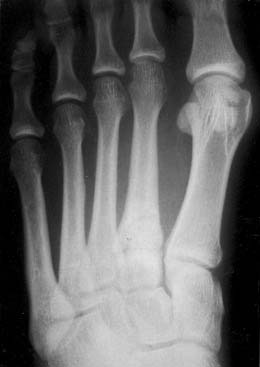
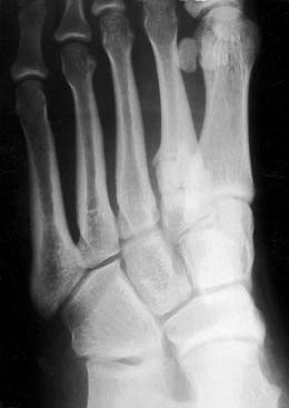
Figure 4-3 An oblique radiograph shows a chronic stress fracture of the base of the second metatarsal.
Related posts:
 Impingement Syndromes of the Foot and Ankle
 Ankle Sprains, Ankle Instability, and Syndesmosis Injuries
 Disorders of the Subtalar Joint, Including Subtalar Sprains and Tarsal Coalitions
 Assessment and Treatment of the Elite Athlete: Helpful Hints and Pertinent Pearls
 Ankle and Midfoot Fractures and Dislocations
 An International Perspective on the Foot and Ankle in Sports
Stay updated, free articles. Join our Telegram channel

Full access? Get Clinical Tree


