Chapter 28 Principles of Rehabilitation for the Foot and Ankle
Introduction
The foot and ankle often are injured during sporting events, recreational activities, and occupational accidents. Injuries to the foot and ankle may be acute or chronic in nature and often cause considerable disability in athletes. Garrick and Requa1 reported that foot and ankle injuries represented more than 25% of the 1600 athletic injuries in their series.2,3 It has been suggested that the sprained ankle is the single most common injury in sports.2,4–7
The foot and ankle serve as the junction of the body to the weight-bearing surface. This elegant collection of tissues, each with a variety of specialized functions, allows efficient, upright stance and locomotion.8 Athletic populations have unique and strenuous demands. Even with minor injuries, improper or incomplete rehabilitation can lead to significant impairment. A detailed, focused approach to rehabilitation of the foot and ankle is crucial to the athlete. Fortunately, most competitive athletes have access to daily evaluation and monitoring of progress, as well as skilled assistance to help them comply with rehabilitation protocols. Recent technologic and procedural advances contribute greatly to the treatment of the competitive athlete. Principles of rehabilitation must continue to advance and keep up to date with technologic and procedural advances. A proper and advanced approach to rehabilitation can provide an environment conducive to a complete, full, and functional recovery.
Cryotherapy/Rest, Ice, Compression, and Elevation (RICE)
Initial treatment of acute foot and ankle injuries and postoperative ankles still follows the RICE principle. There are several cold agents to choose from, including the cold pack, ice bags, cold whirlpool, ice immersion, and the Aircast Cryocuff. The primary objective of ice is to reduce swelling and help manage pain. It has been found that pain is inhibited by cold through a decrease in nerve conduction velocity. As the temperature decreases, there is a corresponding decrease in sensory and motor nerve velocity, eventually causing synaptic transmission to be blocked.9 In our experience, we have found the ankle and foot Cryocuffs to be effective because they combine compression and cold. In addition, elevation can help to reduce hydrostatic pressure and diminish edema. Physiologically, the application of cold agents also results in arteriolar vasoconstriction, a decrease in local metabolism, and an elevation in pain threshold.
The application of cold is most effective immediately after injury or within the first 72 hours. Hocutt et al.10 found that patients with grade III ankle sprains that were treated with ice in the first day returned to functional activities such as running and jumping after 6 days, whereas those treated on the second day went 11 days before they could run or jump. In contrast, those who received heat in the first day had a recovery time of 14.8 days.
A contraindication to cryotherapy is individuals with hypersensitivity to cold. Cold should be avoided in patients with Raynaud’s syndrome or peripheral vascular disease (see Chapter 10). Cold therapy also must be monitored closely in postoperative patients who have wet dressings because the combination of wet dressings with cold application can decrease the skin temperature to a dangerous level.
Range of Motion/Mobilization
Application of early motion on ligament healing demonstrates that the ligament hypertrophies to compensate for decreased tensile strength of the individual fibers. Obviously the amount of tension and stress must not overcome the ultimate load to failure of the tissue and must not lead to fatigue or plastic deformation. Wolff’s law also may apply to these soft tissues, and physiologic stress may allow more functional and stronger healing of soft tissues. Experimental studies of ligaments after injury indicate that exercise and joint motion stimulate healing and influence the strength of ligaments after injury.11–16
Some of the early research on restoration of early range of motion was performed in the hand and the knee. These historical papers revealed insight on how early range of motion decreases complications and actually enhances the healing process. Early mobilization may result in an earlier return to work and daily activity, less muscle atrophy, and better mobility compared with immobilization by casting.14,17,18 The value and benefit of early motion was investigated in the area of rehabilitation after flexor tendon repairs of the hand. The obvious need for full motion in the hand prompted investigation into safe rehabilitation practices, which would eliminate postoperative adhesions and stiffness but allow reliable healing of the tendon. Gelberman et al.19,20 noted an improved healing response, improved strength, and a more normal pattern of vascularity to the healing tendon with protective early mobilization. Several other studies also noted that early range of motion decreased adhesions around the repaired tendon and had a positive influence to the healing tissue.21,22 Early motion after flexor tendon repair has become standard today.
Over the past 2 decades, there have been significant studies in the area of rehabilitation after knee injury and surgery. The focus of knee rehabilitation has centered on obtaining full symmetrical range of motion following a knee injury or surgery. Obtaining full knee extension was one of the most important criteria in allowing the anterior cruciate ligament to heal anatomically and yet still avoid a knee flexion contracture. Close observation of patients who were doing well demonstrated that early range of motion was not detrimental to the ligament (and in fact could be advantageous to proper ligament healing/strengthening) while allowing an earlier and safe return to function.23 Early motion and weight bearing led to a significant decrease in muscle atrophy and decreased complications from arthrofibrosis with an earlier return to function.
Robert Salter and associates24 investigated the effect of joint motion on cartilage nutrition. Early continuous passive motion in synovial joints allows and promotes cartilage nutrition and health. Salter et al.24 demonstrated that small cartilage defects actually could heal with continuous motion, further supporting the benefit of motion on articular cartilage nutrition and healing.
Eiff et al.17 used a prospective randomized study to determine which treatment for first-time ankle sprains, early mobilization or immobilization, is more effective. They reported that, in first-time lateral ankle sprains, although both immobilization and early mobilization prevent late residual symptoms and ankle instability, early mobilization allows earlier return to work and may be more comfortable for patients. Active and passive range of motion is useful to regain motion in cardinal and diagonal planes. Passive range of motion allows the muscles to relax while working the mobility of the joint. Active range of motion requires independent muscle action and incorporates muscle re-education. It is important to work range of motion in the direction opposite of the mechanism of injury (i.e., we allow dorsiflexion and eversion and avoid plantarflexion and inversion initially after a grade II or III lateral ankle sprain). Once the injury has healed, range of motion should include all directions.
In addition to active and passive range of motion, joint mobilization should be incorporated in the rehabilitation program. Accessory movements, termed joint play, are not volitional but accompany voluntary movements or occur passively in response to the ground or other forces. The amount of joint play is a function of ligament and soft-tissue compliance as well as bony configuration.25 Mobilization techniques involve oscillation, distraction, and gliding movements of the joints in the planes of accessory motions. The range of mobilization is always advanced in a graded manner but always stays within the physiologic limits of the joint.25
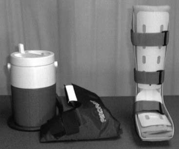
There is much discussion with regard to immediate, short-term protection of ankle injuries. Some of the more common methods consist of elastic wrapping, taping/strapping, semirigid pneumatic ankle brace, nonrigid functional ankle brace, and a removable walking boot. A device we like is the Aircast walking boot with built-in Aircast Cryocuff (Fig. 28-1). The device allows patients to weight bear immediately, work on range of motion by removing the boot, and use a continuous cold/compression device. Once the ankle has healed, a more functional brace is used for return to activity (2-4 weeks after injury). We particularly stress the use of the boot at night for the first 3 to 4 weeks to keep the foot and ankle complex in a 90-degree dorsiflexed position during sleep, when the relaxation of muscular control and the forces on the heel passively place the complex in a plantarflexed and inverted position. The rigid boot counteracts this position.
Protected Weight Bearing
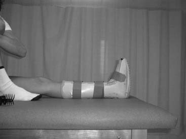
Early weight bearing has been shown to increase the stability of the lateral ankle ligaments after injury while decreasing the amount of muscle atrophy. Protected weight bearing provides a safe and earlier return to activity when appropriate by decreasing joint stiffness, muscular strength deficits, and proprioception dysfunction (Fig. 28-2). We favor a postoperative protocol that allows for early weight bearing whenever possible. We recognize there are times when this is not possible such as in hindfoot fusions. However, in the sports population, early weight bearing can have such a positive impact that we try to tailor our surgical and nonoperative approach to allow early protected weight bearing.
An intriguing area of research that is revealing to us is the investigation of weightlessness. Costill et al.26 examined the effect of a 17-day space flight (essentially, total weightlessness) on muscle. They reported that there was an 11% decrease in peak muscle power, a decrease in muscle fiber diameter, and a 21% decrease in force when the muscle was contracted at peak power velocity. More specifically, Costill et al.26 examined single muscle fiber changes after weightlessness. The single fiber diameter decreases were 20% after 17 days suspended leg weightlessness (for example crutch-assisted nonweight bearing) and demonstrated similar profound muscular atrophy.
Research suggests that early loading of damaged soft tissue can enhance collagen fiber realignment and healing.13,14,16,27,28 Using a removable Aircast walking boot allows the patient to progress to weight bear immediately after injury. Being in a walking boot instead of an ankle cast allows the patient to take the boot off to begin rehabilitation activities. The walking boot provides more support than elastic wrapping, taping, and other semirigid bracing systems, and it also allows the patient the ability to apply cold compression simultaneously.
Gait Evaluation
Normal gait is composed of two phases, a stance phase (60%) and a swing phase (40%). The stance phase is composed of five categories, including initial contact (heel strike), loading response (foot flat), midstance (single leg support), terminal stance (heel off), and pre-swing (toe-off). The swing phase consists of initial swing (acceleration), midswing, and terminal swing (deceleration).29–31
Proprioception
Many rehabilitation programs often fail to pay attention to proprioception deficits. Proprioception is the ability of the body to vary the forces of muscles in response to outside forces. Muscles, tendons, and joint receptors provide this information, which affects posture, muscle tone, kinesthetic awareness, and coordination.29,30 When an individual is injured, the proprioceptive input to that joint is altered and diminished. Diminished proprioception can lead to a recurrence of injury because of the joint’s decreased ability to respond to outside forces.
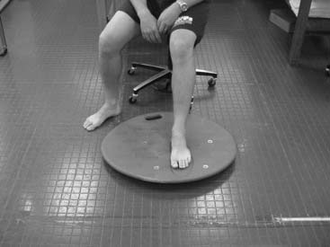
Proprioception can be improved with a number of treatment techniques. Early weight bearing can help to decrease the amount of proprioception loss. A patient can practice standing with equal weight on both feet, progressing to single leg stance. A biomechanical ankle proprioception system (BAPS) board or kinesthetic awareness trainer (KAT) can be used as a patient advances through rehabilitation (Fig. 28-3).
Cardiovascular Activities
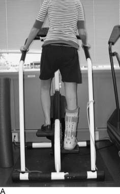
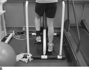
Our experience with and observation of clinical healing and postoperative wound healing have proven that it is important to progress the patient’s activity gradually. Increasing the time increments of 10 minutes a week on a bike will allow the patient to be working approximately 30 minutes per session in a 3-week span (Table 28-1). Typically, low-impact, weight-bearing exercise will be introduced when the athlete is able to walk normally in a protective device and regular shoe. The rehabilitation program will begin replacing one day of bike with a StairMaster/elliptical machine (Fig. 28-4, A and B). We allow an additional day of StairMaster or elliptical each successive week until the athlete has been converted to StairMaster or elliptical 4 to 6 days per week. The athlete will continue to increase low-impact, weight-bearing exercise as tolerated. We have found that when an athlete can work out on the StairMaster or elliptical machine 4 to 5 days a week for 30-plus minutes, it is safe to initiate running. Running should gradually replace StairMaster/elliptical each week. It is important to give the athlete a set of running guidelines that allows for a gradual progression of activity (Table 28-2).
Table 28-1 Increase Exercise Capacity Program (with boot/postoperative shoe/brace on)
 Exercise 10 minutes on a stationary bike 3 days a week. Exercise 10 minutes on a stationary bike 3 days a week. |
 Exercise 20 minutes on a stationary bike 4 days a week. Exercise 20 minutes on a stationary bike 4 days a week. |
 Exercise 30 minutes on a stationary bike 4 days a week. Exercise 30 minutes on a stationary bike 4 days a week. |
| •Once you are able to ride the bike 30 minutes a day for 4 days a week, then you may start replacing one of your days of biking per week with 1 day of StairMaster or elliptical trainer. You will do the StairMaster or elliptical for the same amount of time you normally would ride the bike. |
< div class='tao-gold-member'>
Related posts:
 Impingement Syndromes of the Foot and Ankle
Stress Fractures: Their Causes and Principles of Treatment
Disorders of the Subtalar Joint, Including Subtalar Sprains and Tarsal Coalitions
Foot and Ankle Injuries in Dancers
Diagnostic and Operative Ankle and Subtalar Joint Arthroscopy
Lesser-Toe Disorders
Impingement Syndromes of the Foot and Ankle
Stress Fractures: Their Causes and Principles of Treatment
Disorders of the Subtalar Joint, Including Subtalar Sprains and Tarsal Coalitions
Foot and Ankle Injuries in Dancers
Diagnostic and Operative Ankle and Subtalar Joint Arthroscopy
Lesser-Toe Disorders
Stay updated, free articles. Join our Telegram channel

Full access? Get Clinical Tree


