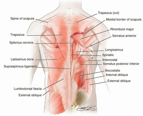
Muscle |
Origin |
Insertion |
Innervation |
Blood Supply |
Superficial layer |
Trapezius |
Medial third of superior nuchal line of occiput, external occipital protuberance, and ligamentum nuchae; spinous processes of C7-T12 |
Lateral third of clavicle, acromion, spine of scapula |
Motor supply from spinal accessory nerve, sensory fibers from C3 to C4 |
Transverse cervical artery |
Latissimus dorsi |
Spinous processes of T7—sacrum, medial third of iliac crest, ribs 9-12, inferior angle of scapula |
Floor of bicipital groove |
Thoracodorsal nerve (C7, C8) |
Thoracodorsal artery |
Levator scapulae |
Transverse processes of C1-C4 |
Medial border of scapula |
Dorsal scapular nerve (C5), with branches of C3-C4 innervating upper part of muscle |
Dorsal scapular artery |
Rhomboid major |
Spinous processes of T2-T5 |
Medial border of scapula |
Dorsal scapular nerve (C5) |
Dorsal scapular artery |
Rhomboid minor |
Caudal end of ligamentum nuchae, spinous processes of C7-T1 |
Medial border of scapula |
Dorsal scapular nerve (C5) |
Dorsal scapular artery |
Intermediate layer |
Serratus posterior superior |
Spinous processes of C7-T3 |
Ribs 1-4 |
Intercostal nerves |
Posterior intercostals arteries of T1-T4 |
Serratus posterior inferior |
Thoracolumbar fascia, spinous processes of T11-L2 |
Ribs 9-12 |
Intercostal nerves |
Posterior intercostal arteries, subcostal artery, and L1-L2 lumbar arteries |
Levatores costarum |
Tip of transverse process of C7-T11 vertebrae |
Rib below level of origin |
Posterior rami of thoracic spinal nerves |
Dorsal intercostal arteries |
Deep layer |
Erector spinae (vertically oriented and superficial) |
Iliocostalis Longissimus Spinalis |
Iliac crest, sacrum, transverse and spinous processes of vertebrae, and supraspinal ligament |
Ribs, transverse and spinous processes of vertebrae, posterior aspect of skull |
Segmental innervation by dorsal primary rami of spinal nerves C1-S5 |
Segmental supply by deep cervical arteries, posterior intercostal arteries, subcostal artery, and lumbar arteries |
Transversospinalis (obliquely oriented and intermediate) |
Semispinalis |
Transverse processes T1-T12 |
Spinous processes of C2-T5 |
Dorsal rami of spinal nerves |
Segmental arteries from aorta |
Multifi |
dus Articular processes of cervical vertebrae, transverse processes of thoracic vertebrae, mammillary processes of lumbar vertebrae, posterior superior iliac spine |
Spinous processes of C2-L5 |
Dorsal rami of spinal nerves |
Segmental branches from aorta |
Rotatores |
Transverse processes |
Base of spinous processes above Long skip one level; short attach at level above |
Dorsal rami of spinal nerves |
Segmental branches from aorta |
Deepest muscle |
Interspinales |
Spinous processes |
Spinous processes one level above |
Dorsal rami of spinal nerves |
Segmental branches from aorta |
Intertransversarii |
Anterior and posterior transverse processes of cervical vertebrae, transverse and mammillary processes of lumbar vertebrae |
Anterior and posterior processes of cervical vertebrae one level above, transverse and accessory processes of lumber vertebrae one level above |
Dorsal rami of spinal nerves |
Segmental branches from aorta |


