Minimally Invasive Lumbar Microdiscectomy and Laminectomy
David Greg Anderson
Christopher K. Kepler
Victor M. Popov
DEFINITION
Lumbar microdiscectomy is the most common spinal operation.1
Lumbar spinal stenosis is the most common indication for spinal surgery in elderly patients.1
The pathology of lumbar spinal stenosis is a combination of degenerative changes involving the disc space, facet joints, and ligamentum flavum, which ultimately leads to compression of the neural elements and, in some cases, neurogenic symptoms.6
Studies have shown favorable outcomes from surgery for patients with herniated disc disease and lumbar stenosis.1,2,12
Minimally invasive surgical decompression has been shown to decrease perioperative morbidity and quicken patient recovery.3,6,7
This chapter reviews the technique of performing lumbar decompressions for herniated disc disease and lumbar stenosis using a minimally invasive approach.
HISTORY AND PHYSICAL FINDINGS
Although the exact clinical presentation varies from person to person, most patients with herniated disc disease and lumbar stenosis present with pain radiating into the lower extremities.
The classic presentation of herniated disc disease involves pain radiating into a single extremity along a specific dermatomal distribution. There are often associated neurologic findings, including changes in strength, sensation, and reflexes.
The classic presentation of lumbar stenosis is neurogenic claudication, which involves crampy pain radiating into one or both extremities when standing and walking. The pain commonly progresses from proximal to distal and is improved or relieved by spinal flexion (ie, leaning forward or sitting down).
This is in contrast to vascular claudication, which is relieved by standing still. Significant neurologic deficits in lumbar stenosis are uncommon; however, those with herniated disc disease will commonly show changes in reflexes, motor, and sensory functioning. Acute loss of bowel or bladder control in the setting of significant compression of the cauda equina is a surgical emergency.
Nonsurgical measures that may be considered for both herniated disc disease and lumbar stenosis include nonsteroidal anti-inflammatory drugs, epidural steroids, and physical therapy.
SURGICAL MANAGEMENT
Preoperative Planning
Surgical intervention may be considered for patients with severe, ongoing symptoms of leg pain that are unresponsive to nonsurgical therapy.
It is important to demonstrate anatomic compression of lumbar nerve roots in a distribution that correlates to the clinical symptoms. This is usually done with either magnetic resonance imaging (MRI) or computed tomography (CT) myelography.
Positioning
The procedure is typically done under general anesthesia, although epidural or spinal anesthesia may be used according to surgeon preference.
Prior to surgery, prophylactic antibiotics and lower extremity compression stockings are administered.
The patient is positioned prone on a spinal frame, which allows fluoroscopic imaging of the spine (FIG 1).
Care should be taken to ensure no compression of the abdominal region.
Care should be taken to ensure accessibility for fluoroscopic imaging.
A standard sterile preparation and drape of the lumbar region is performed.
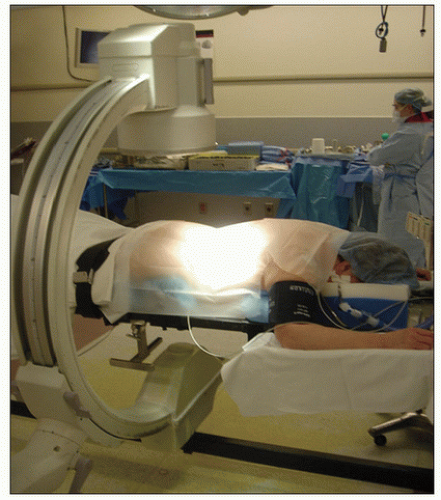 FIG 1 • Positioning of the patient in the prone position on a radiolucent operative table. |
TECHNIQUES
▪ Unilateral Decompression
Incision
Palpable landmarks, including posterior superior iliac spine, intercrestal line, and spinous processes, are demarcated and used to determine the approximate level for the skin incision.
A spinal needle is introduced lateral to the midline in the proposed location of the surgical incision and directed to avoid penetration of the spinal canal (to minimize inadvertent penetration of the dura) (TECH FIG 1).
The C-arm is used to obtain a lateral projection of the spine and spinal needle so the location and trajectory of the needle can be used to plan the incision.
A skin incision equal in length to the diameter of the tubular retractor is made through the skin and underlying fascia. The incision is placed lateral to the spinous process.
Placement of the Tubular Retractor
In cases of herniated disc disease, the incision is made on the side of the herniation.
In cases of lumbar stenosis, the incision is positioned on the side that will give the best access to both sides of the spinal canal or will allow decompression of the region of symptomatic stenosis without sacrificing excessive facet joint or overthinning the pars interarticularis region.
For ipsilateral decompression, the skin incision is generally positioned about 1.5 to 2 cm from the midline.
For bilateral decompression, the skin incision is generally positioned 3 to 4 cm lateral to the midline to allow angulation of the tubular retractor across the midline to reach the contralateral side of the spinal canal.
A small Cobb elevator is used to elevate the periosteum at the site of the operative exposure (TECH FIG 2A). We prefer to use this rather than dilation over a K-wire, which has the risk of penetrating the dura and is less effective in creating the working space adjacent to the lamina.
Dilators are placed through the incision to expand the operative portal (TECH FIG 2B).
A tubular retractor of appropriate length is selected and placed over the dilators to the level of the spine (TECH FIG 2C).
The diameter of the tubular retractor used depends on the nature of the planned surgical procedure. As a general rule, a 14- to 18-mm diameter tubular retractor is used for a microdiscectomy procedure, whereas an 18- to 20-mm tubular retractor is used for spinal stenosis cases.
The tubular retractor is secured to the table-mounted retractor holder, and the position of the retractor is verified with lateral fluoroscopy (TECH FIG 2D).Related posts:
 Occipitocervical and C1-C2 Fusion with Instrumentation
Transforaminal and Posterior Lumbar Interbody Fusion
Primary and Metastatic Tumors of the Spine: Total En Bloc Spondylectomy
Vertebral Column Resection for Severe Rigid Spinal Deformity through an All Posterior Approach
Anterior Interbody Arthrodesis with Instrumentation for Scoliosis
Posterior Cervical Approach
Occipitocervical and C1-C2 Fusion with Instrumentation
Transforaminal and Posterior Lumbar Interbody Fusion
Primary and Metastatic Tumors of the Spine: Total En Bloc Spondylectomy
Vertebral Column Resection for Severe Rigid Spinal Deformity through an All Posterior Approach
Anterior Interbody Arthrodesis with Instrumentation for Scoliosis
Posterior Cervical Approach

Stay updated, free articles. Join our Telegram channel

Full access? Get Clinical Tree


