pigment deposition

Chapter 12 Abnormal pigment deposition


Haemosiderin


Haemosiderin is a golden-brown pigment derived from the breakdown of haemoglobin. It can be demonstrated in tissue sections by the Prussian blue reaction, which is utilized in histological stains such as Perls stain. Small amounts are normal in the bone marrow, but otherwise its presence is usually pathological.


Localized haemosiderin deposition occurs in inflamed tissues from the breakdown of haemoglobin in extravasated red blood cells. The haemosiderin is taken up by macrophages (Fig. 3.12.1). The presence of this pigment is used by pathologists as an indicator that inflammation has occurred, and it may be mentioned in histopathology reports for this reason.
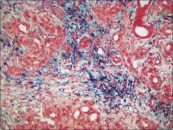


If there is systemic iron overload, haemosiderin is deposited in many organs and tissues. Causes include haemolytic anaemia, recurrent blood transfusions, increased absorption of iron from the intestine, and inherited disorders of iron metabolism. The most common inherited disorder of iron metabolism is a homozygous recessive disorder called hereditary haemochromatosis and caused by a mutation of the gene HFE. The consequence is failure of the normal regulation of iron absorption from the intestine so that excessive quantities of iron are absorbed from the gut lumen. Large amounts of iron in the liver, heart, pancreas and skin can cause hepatic fibrosis, heart failure, diabetes and skin pigmentation, respectively.


Pathologists tend to use the term haemosiderosis for the presence of iron in the tissues, while the disease caused by the iron is called haemochromatosis.

< div class='tao-gold-member'>

Stay updated, free articles. Join our Telegram channel

Jul 3, 2016 | Posted by in MUSCULOSKELETAL MEDICINE | Comments Off on pigment deposition

Full access? Get Clinical Tree

Get Clinical Tree app for offline access