Fig. 45.1
Benign PMT. MRI showing a high signal intensity juxtacortical heterogeneous mass in the left femur in a coronal view
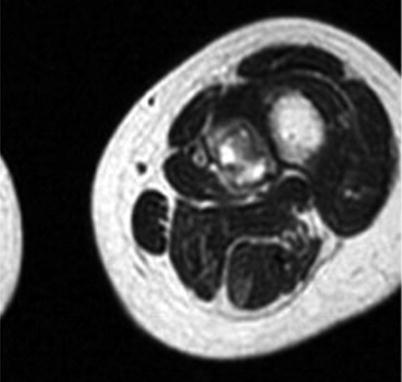
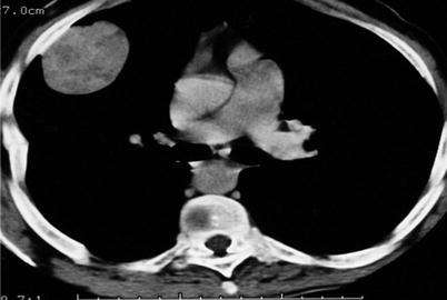
Fig. 45.3
Benign PMT. CT shows an ovoid lesion attached to the internal wall of the rib cage. There is no mineralized area
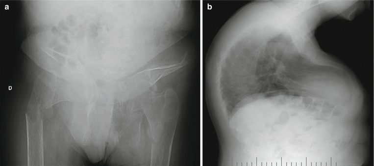
Fig. 45.4
Same case as in Fig. 45.1. Radiographs show diffuse, intense osteopenia, a deformed pelvis, and insufficiency fractures of both femurs (a) and intense deformities of the spine and the rib cage (b). Bone consistency got back to normal, and fractures healed after removal of tumor
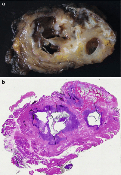
Fig. 45.5
Same case as in Fig. 45.1. The specimen contained cysts surrounded by tan soft tissue with white areas of ossification (a). (b) A whole specimen histological mount; cellular areas are seen around the cysts with peripheral ossification
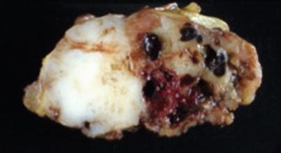
Fig. 45.6
Another slab section of the same case as in Fig. 45.2. White, fish-flesh-like areas denounce cellular areas of lesion. Hemorrhagic and tan soft areas are also seen
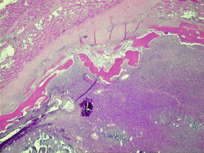
Fig. 45.7
PMTMCT – peripheral ossification is seen between a fibrous capsule and cellular area of the tumor
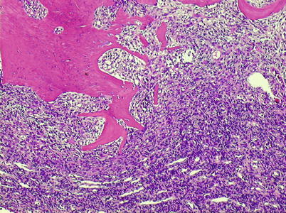
Fig. 45.8
PMTMCT – richly vascularized tumor tissue, hemangiopericytoma-like pattern
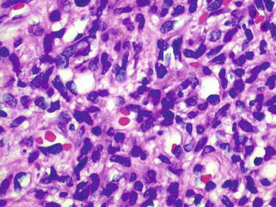
Fig. 45.9
PMTMCT – numerous capillaries in a high proliferation of bland, spindled to stellate cells
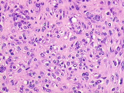
Fig. 45.10
PMTMCT – area with background resembling chondroid matrix
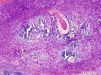
Fig. 45.11
PMTMCT – low-power view of area with peculiar calcifications, an unusual “smudgy” matrix that calcifies in a characteristic flocculent fashion
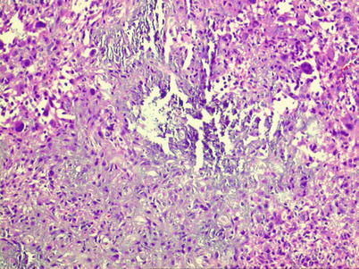
Fig. 45.12


PMTMCT – higher-power view of focus of flocculent calcifications
Related posts:


Stay updated, free articles. Join our Telegram channel
Full access? Get Clinical Tree
