Fig. 52.1
(a) Roentgenogram showing an elongated area of cortical irregularity along the medial cortical tibia. (b) CT scan shows an intracortical small lytic lesion. (c) MRI, the arrow indicates the area of cortical erosion

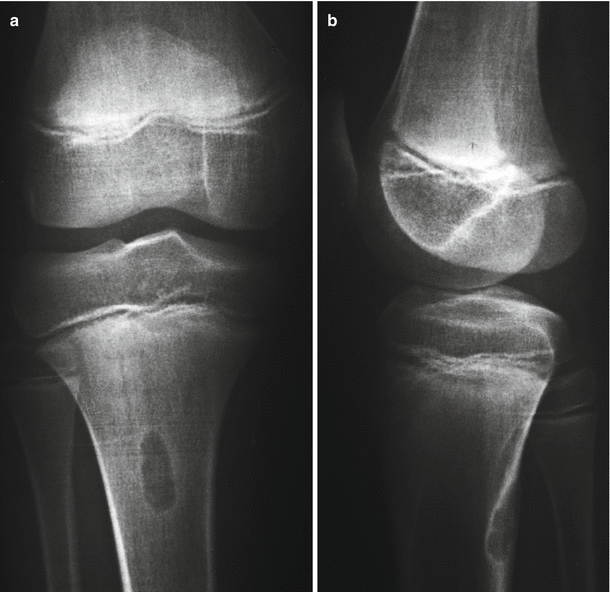
Fig. 52.2


(a, b) Anteroposterior and lateral X-ray of a periosteal desmoid in the posterior cortex of tibial metaphyseal area
Stay updated, free articles. Join our Telegram channel

Full access? Get Clinical Tree


