Fig. 18.1
Radiograph of the proximal femur revealing cortical erosion (saucerization) along the medial margin. Inferior margin of the lesion shows a cortical beak
Chondroid calcifications are observed in about half the tumors, and some show extensive calcification.
Even when they do not have any chondroid calcification, the shape of the cortical defect suggests the diagnosis. The tumor does not involve the medullary cavity.
CT Features
CT shows detailed calcification and saucerlike cortical erosion of the bone. Cortical scalloping with variable degree of sclerosis is seen related to buttresses of periosteal new bone.
MRI Features
On MR imaging, tumor shows low to intermediate signal intensity on the T1-weighted images and high signal intensity on the T2-weighted image. Calcification and matrix mineralization of the tumor appear as decreased signal intensity areas on all pulse sequences.
Images with contrast enhancement show variable findings. However, in general, the tumor shows the peripheral and septal pattern enhancement of cartilage lesions.
Image Differential Diagnosis
Periosteal Chondrosarcoma
The most reliable diagnostic criterion would be the size of the lesion. Malignancy should be suspected when bigger than 3 cm. Periosteal chondrosarcomas are usually large (over 5 cm) and show no radiologic evidence of solid periosteal new bone buttressing at the margins, which is a characteristic finding of periosteal chondroma.
Periosteal Osteosarcoma
Periosteal osteosarcoma can be recognized by the feathery perpendicular calcific striae seen on radiographs. Periosteal osteosarcomas lack the characteristic periosteal new bone buttress formation typical of periosteal chondromas and tend to fade into the soft tissues.
Pathology
Gross Features
Grossly, periosteal chondroma is a well-circumscribed, lobulated cartilaginous mass.
The outer surface is covered by fibrous periosteum of the involved bone.
The cortex underneath is indented and thickened. Scalloping and erosion of the underlying cortex may be seen.
The tumors are usually less than 3 cm in greatest dimension. Any surface cartilage lesion larger than 3–5 cm should be suspected to be malignant.
Histological Features
Periosteal chondromas show characteristic lobules of hyaline cartilage (Fig. 18.2).
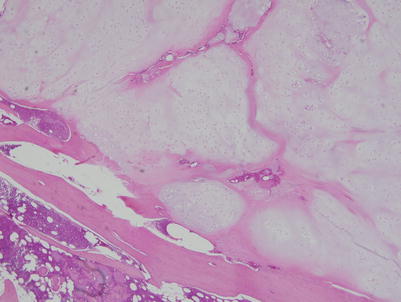
Fig. 18.2
At low-power magnification, the lesion shows characteristic lobular hyaline cartilageRelated posts:

Stay updated, free articles. Join our Telegram channel

Full access? Get Clinical Tree


