Fig. 20.1
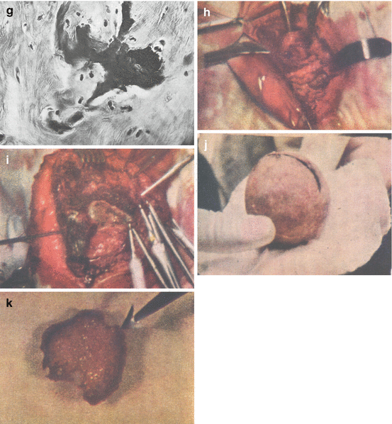
Ochronosis in joint tissues. Histological images of the connective tissue from the tendons and fibrous capsule of a patient with AKU showing deposition of ochronotic pigment (a–g). Photographs of the open hip joint (h), knee joint (i), head of femur (j) and patella (k)
The tough connective tissue of tendons and fibrous capsule contains dark black, macroscopically recognisable foci of pigment of various sizes, from large pigmented areas visible macroscopically to individual pigment granules visible only by microscope. During the processing, the pigment accumulated in large foci falls apart, and during the slicing, it gets to marginal areas adjacent to tough connective tissue. Tough connective tissue around the focus has a characteristic concentric arrangement, and fibres run around pigment granule in several layers. At many places, the tissue loses its regular pattern of collagen fibres, and irregular spaces are filled with fibrous cells with hypertrophic cytoplasm between them or their fascicles arise. The cell bodies are of considerable size, with polygonal or oval shape, and it contains an oval nucleus with well-stainable chromatin structure. The cytoplasm of these cells contains various amounts of brown pigment granules from a few pieces up to large amounts filling the whole cell body and covering the nucleus. It can be seen that cells overfilled with pigment decompose, pigment granules are released from them, and they gradually form to freely deposited bigger or smaller granules and lumps of pigment mass between the fibres (Fig. 20.1a–c).
There are no signs of inflammatory reaction in the connective tissue around such pigment foci. However, adjacent connective tissue is relatively well vascularised, and small blood vessels (small arteries, arterioles and veins) can be seen in divided and loose connective tissue. Adventitia of these vessels contains large amount of polygonal and oval cells with larger or smaller amount of brown pigment granules in cytoplasm. The number of these cells varies; there are only a few of them at some places, but in the other places, they form apparent perivascular foci (Fig. 20.1d), or they can even be present independently from blood vessels, especially in thinner interstitial connective tissue. More distant places from pigment foci usually have normal structure of tough connective tissue with typical nuclei of tendon cells.
In some areas, especially under the surface of tendons and capsule, degenerative changes can be observed in areas containing pigmented cells or even large pigment foci (Fig. 20.1e). Groups of well-demarcated connective tissue fibres of various sizes are hyalinised; their fibrillar structure has been lost creating a homogeneous, deeply basophil mass with sporadically included polygonal cells with pigmented cytoplasm. Even in adjacent areas of such hyalinised parts, cells containing pigment granules can be seen in connective tissue. Signs of necrosis and thin fibrillar structures characterised by intense basophilia can be sporadically seen at some places. The thin coating of joint cartilage is incomplete. Degenerate hyaline cartilage with distinctive fibrillar matrix (Fig. 20.1f) and well-demarcated pigment foci of dark brown colour and various sizes (Fig. 20.1g) can be seen in the sections from greyish spots of this coating. These foci are distinctly separated from adjacent area; their margins look like they have been cut, while there are no particular reactive signs in adjacent tissue.
Some specific data (situation from open hip joint can be seen at Fig. 20.1h) have to be pointed out from the surgical report of our 70-year-old patient dated January 24, 1953: supraacetabular insertion of the rectus femoris muscle as well as acetabular and trochanteric insertion of the capsule had marked dark blue colouration. It results from the second surgical report dated June 20, 1953, that the subcutaneous connective tissue of the right knee joint as well as deep muscular fasciae had dark blue colouration. The ligamentum patellae proprium and the whole tendon of the quadriceps femoris muscle were massively impregnated by ochronotic pigment (Fig. 20.1i). On the other hand, the medial femoral condyle was also covered by dark blue pigment, and it impregnated the whole absolutely damaged cartilaginous and bone part of the medial and lateral condyle.
The following changes were found on the femur head and patella during their surgical removal:
Femur head: it did not have any cartilaginous glossy coating except some small remains. The epiphysis surface had grey colouration, and it was dull. In the lateral quadrant of the epiphysis, there was one 2.5 cm long, not a completely continuous line that looked like artificially drawn by carbon pencil (Fig. 20.1j). Not far from it, there was another 5 cm long arch-like line. The subchondral part and spongy bone were markedly sclerotic. Section through the head showed that these pigment lines reached up only 4–6 mm in subchondral direction.
The articular facet of the patella was bended, rugged and thickened. There was a nearly complete continuous pigment line of 3–6 mm width going through the peripheral part (Fig. 20.1k). At the place where the tendon of the quadriceps femoris muscle goes to the patella, carbon-coloured deposits were visible. After splitting the patella, we could see that pigment line reaches only subchondrally and does not reach deeper layer of the spongy bone. It was a completely identical picture as on resected proximal epiphysis of the femur. It has to be noted that neither in the acetabulum nor in the intra-articular part of the knee joint, no free particles of detached cartilage that would be impregnated by ochronotic pigment were found. Multiple foci of ochronotic pigment are located especially in loose fibrous connective tissue. We also found the location of fibrous tissue cells with pigment granules in perivascular area as well as pigment foci among the fibres of solid fibrous connective tissue. Solid fibrous tissue showed the signs of hyaline degeneration, and it was pigmented. ‘Shadows’ of chondrocytes can be seen in the mass of pigment indicating that ochronosis occurred by uptake of pigment into cartilage matrix and chondrocytes and/or trapping of chondrocytes in the matrix during the course of pigmentation. The structure of undamaged cartilage areas shows typical senile changes with multiple isogenic groups of cells.
Stay updated, free articles. Join our Telegram channel

Full access? Get Clinical Tree


