Fig. 45.1
Dejour’s trochlear dysplasia classification. Type A, crossing sign, trochlear normal (>145°); Type B, crossing sign, supratrochlear spur, flat or convex trochlea; Type C. crossing sign, double contour; Type D, crossing sign, supratrochlear spur, double contour [11]
The patellar height can be measured with the Insall-Salvati index or Caton-Deschamps index. The first is the ratio of the length of the patellar tendon to the longest sagittal diameter of the patella [12]. The second is the ratio of the distance from the lower edge of the articular surface of the patella to the anterosuperior angle of the tibia outline [13]. Two basic types of axial views (Merchant and Laurin) can be done with different types of measurement. The Merchant can measure the sulcus angle and the congruence angle [14]. The Laurin can measure the lateral patellofemoral angle and the patellofemoral index [15]. There is a great variability in the patellar shape, and Wiberg classified them into three types. The most frequent type of patella in patellar dislocation is Wiberg type II [16].
Use of CT scan imaging for exploration of the patellofemoral relationship has led to a better understanding of the dynamics of this joint in normal and pathologic knees, and the use of it is widely accepted. It allows the study of many keen parameters. The tibial tubercle-trochlear groove distance (TT-TG) is the instrumental measurement of the “Q” angle. First described in 1978 by Goutallier as radiologic measurement, then in 1987 H. Dejour adapted this measurement to the CT scan. We use two cuts to measure the distance between the central point of the tibial tubercle (TT) and the deepest point of the trochlear groove (TG). Normal value is around 12 mm and in the patellar dislocation population is greater than 20 mm [10]. This pathologic value can be indicative for tibial tubercle medicalization. The patellar tilt is the angle between the transverse axis of the patella and the posterior femoral condyles. Eighty-three percent of the patellar dislocation population has a value greater than 20° compared to 3 % in the normal group [10]. We can obtain and even measure femoral anteversion and external tibial rotation. MRI has been considered more useful in defining cartilage status than measuring patellofemoral instability parameters. Some authors have compared, with the same reproducibility, TT-TG measurement with CT scan and MRI, but still need more study [17].
45.4 Treatment Strategy
Surgical treatment of patellar instability has two approaches. One is that anatomic alignment is the most important factor, and all “malalignment” factors must be correct. The other approach is softer because it recognized the importance of soft tissue restraint to lateral patellar translation: to stabilize or recreate, this restraint can be sufficient in many cases with bony defects uncorrected [18]. The first evaluation is about conservative versus surgical treatment. Surgical treatments contain soft tissue and bony procedures. Soft tissue procedures: lateral release, medial soft tissue reefing, proximal realignment, and MPFL reconstruction. Bone procedures are distal realignment, trochleoplasty, and distal femoral osteotomy. Substantial controversy exists about treatment strategy. From a logical standpoint, the procedures adopted should correct the observed root abnormalities, and it is more likely that a combination of procedures would correct those abnormalities one by one, rather than one standard procedure for every case. To remedy patellar instability, the surgeon will need to combine soft tissue and bony procedures to address all involved factors, each corrected individually.
For acute first-time dislocation, the classic treatment is conservative. The more important exception to this is the presence of an osteochondral fracture. Some authors propose acute repair in cases of substantial medial structure disruption and a laterally subluxated patella with a normally aligned opposite knee. The main goals of conservative treatment are swelling and pain remission, as well as restoration of range of motion. Quadriceps strengthening is another goal of the conservative management strategy, and good quadriceps strength seems to alleviate symptoms, but whether it prevents future dislocation is unclear. Immobilization for up to 6 weeks may help medial structure heeling, but stiffness is a problem. If recurrent dislocation occurs, they will put the patient in a different category from treatment purposes: the chronic dislocation group.
45.4.1 Lateral Release
We can find LR treatment isolated or associated procedures. Isolated LR has no role in treatment of acute or recurrent patellar instability because from a mechanical perspective, isolated LR cannot correct the causes of patellar instability. Isolated LR can be a successful procedure in patients with isolated lateral patellar tightness. Associated with medial reefing or to a proximal/distal realignment, we have a better result [19].
45.4.2 Medial Reefing
The indication for arthroscopic medial reefing after acute dislocation is the presence of persistent patellar dislocation or detachment of the medial retinaculum or MPFL from the patellar insertion (usually the lesion is on the femoral side) [3]. A relative contraindication can be trochlear dysplasia grade B or C according to H. Dejour or rupture of MPFL on the femoral insertion.
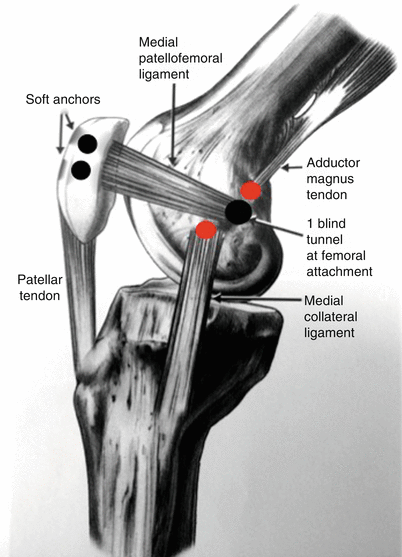
45.4.3 Medial Patellofemoral Ligament Reconstruction
The increased mobility after dislocation appears to be attributable to medial retinacular deficiency. On cadaver it has been shown that the MPFL is the most important structure resisting lateral patellar motion. According to literature MPFL injury may be present in approximately 65 % of cases [20]. Restoration of the MPFL has been described by primary repair alone, repair with augmentation, and reconstruction alone. MPFL repair has been approached in various ways. The repair may be done acutely or after a period of initial healing. Repair has been described at the patellar insertion of the MPFL. Our favorite technique for the reconstruction is with the semitendon fixed to the rotula with two soft anchors and half tunnel in femur with a screw. Previously we create a slot on the medial side of the rotula to allow integration, and we don’t perform any tunnel in the rotula to avoid fractures, especially in small rotula, or cartilage damage. The key point is to choose the right point of insertion to restore proper kinematics. Non-anatomical femur insertion can lead to excessive medial pressure on the rotula cartilage [21]. We measured rotula and selected two-third proximal to implants the two soft anchors. Femur position is the most difficult step of the reconstruction. The MPFL originates from a ridge between the medial epicondyles and the adductor tubercle (Fig. 45.2).


