3 Part 1
Effects of acupuncture needling on connective tissue
Needles and connective tissue
Acupuncture needles are fine, filiform needles used both in traditional acupuncture as well as in more recently developed ‘dry needling’ techniques. An important characteristic of acupuncture needles is that their small diameter (typically less than 300 µm) allows a unique form of interaction to occur between the needle and connective tissue (Langevin et al. 2001a). When acupuncture needles are rotated, collagen bundles adhere to the needle and wind around its shaft, creating a small ‘whorl’ of collagen in the immediate vicinity of the needle, i.e., within a few millimeters (Kimura et al. 1992, Langevin et al. 2002). Winding of collagen, which can occur with as little as half a revolution of the needle, causes connective tissue to follow the rotating needle and then adhere to itself, further increasing the mechanical bond between needle and tissue. Once a needle–tissue bond is established, any further motion of the needle, either rotation or up-and-down, effectively pulls the tissue along the direction of needle motion.
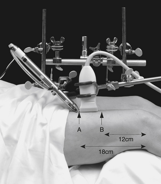
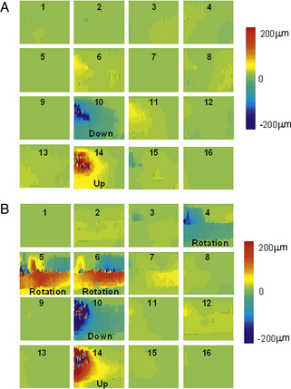
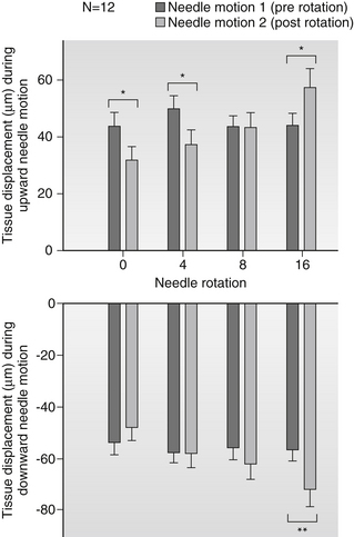
In humans, winding of connective tissue is quantified using robotic acupuncture needling as well as ultrasound elastography imaging techniques (Figure 3.1; Langevin et al. 2001b, 2004). Both uni-directional, whereby the needle is rotated continuously in one direction, and bi-directional rotation, with the needle rotated back and forth, cause a measurable increase in the amount of force necessary to pull the needle out of the skin (Langevin et al. 2001b). With uni-directional rotation, the torque developing at the needle–tissue interface increases exponentially as the needle rotates. This phenomenon is similar to the exponential increase in friction forces that develops as a cable is wound around a winch drum. With bi-directional rotation, needle torque gradually increases over successive rotation cycles. This is likely due to incomplete unwinding of tissue at the end of each cycle, leading to a gradual build-up of torque in the tissue. Thus, both uni-directional and bi-directional needle rotation can cause the tissue to ‘grip’ the needle. Even small amounts of needle motion can cause measurable tissue displacement up to several centimeters away from the needle (Figure 3.2). Furthermore, increasing the amount of rotation causes a linear increase in the amount of tissue displacement during subsequent axial needle motion (Figure 3.3; Langevin et al. 2004). Acupuncture needles therefore can be used as ‘tools’ to cause specific mechanical stimulation of connective tissue that can be quantified in vivo in humans and animals.

Figure 3.1 Method used for ultrasound elasticity imaging during robotic acupuncture needling
An acupuncture needle is inserted and manipulated using a robotic acupuncture needling instrument and an ultrasound cine-recording is acquired during the needle manipulation.
Reprinted with permission from Langevin HM, Konofagou EE, Badger GJ et al. (2004) Tissue displacements during acupuncture using ultrasound elastography techniques. Ultrasound Med Biol 30: 1173-1183.

Figure 3.2 Example of tissue displacement maps obtained by post processing of sequential ultrasound images acquired during acupuncture needle manipulation
Gray scale values indicate upward or downward tissue displacement during (A) downward and upward needle motion and (B) rotation followed by downward and upward needle motion.
Reprinted with permission from Langevin HM, Konofagou EE, Badger GJ et al. (2004) Tissue displacements during acupuncture using ultrasound elastography techniques. Ultrasound Med Biol 30: 1173-1183

Figure 3.3 Tissue displacement during upward and downward needle motion before (dark grey bars) and after (light grey bars) increasing amounts of needle rotation
After needle rotation, there was a significant (p<0.001) linear increase in tissue displacement as a function of needle rotation for both upward and downward needle motion.
Reprinted with permission from Langevin HM, Konofagou EE, Badger GJ et al. (2004) Tissue displacements during acupuncture using ultrasound elastography techniques. Ultrasound Med Biol 30: 1173-1183
Stay updated, free articles. Join our Telegram channel

Full access? Get Clinical Tree


