Fig. 8.1
(a) Lateral x-ray of the skull. Note the oval sclerotic lesion in the temporoparietal area. (b, c) CT scan shows a dense homogeneous lesion protruding from the outer table. The diploe and the inner table are not involved

Fig. 8.2
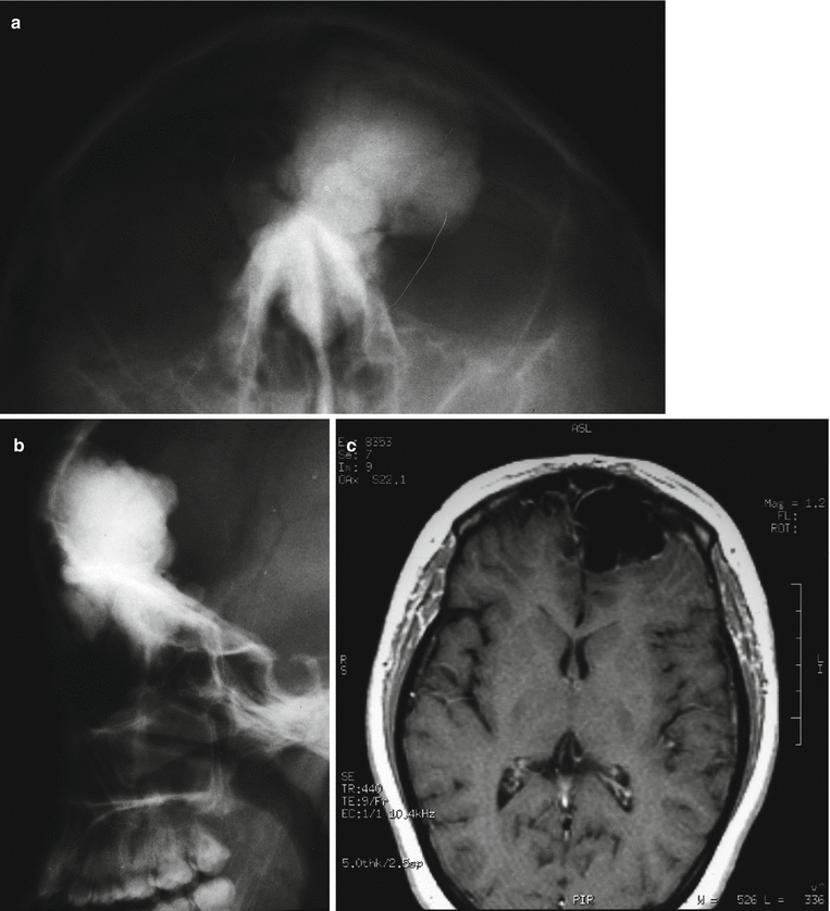
(a, b) X-ray showing a homogeneous sclerotic mass in the frontal bone of the skull. (c) MRI clearly shows a parosteal osteoma arising in the inner table of the skull projecting into the frontal lobe

Fig. 8.3


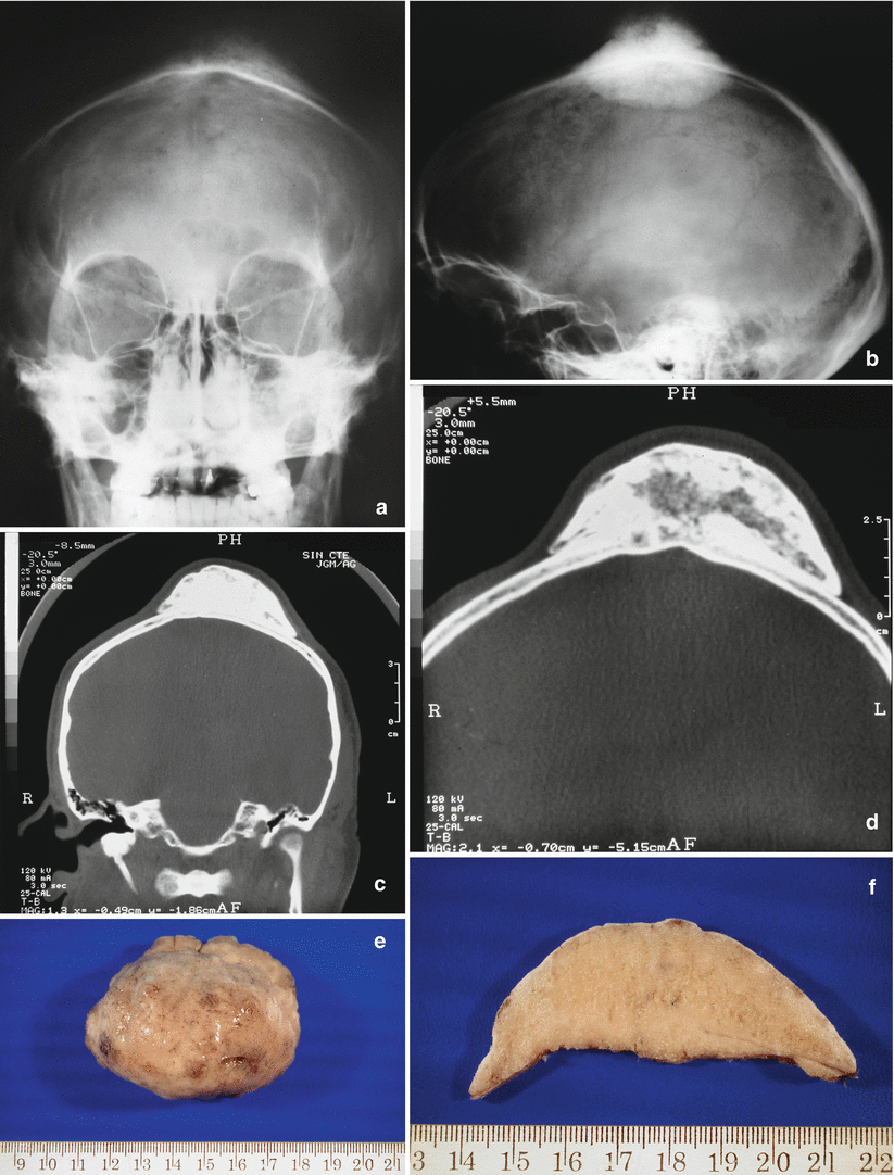
(a, b) Anteroposterior and lateral x-ray of the skull. Note a surface radiodense mass. (c, d) Coronal and axial CT scan clearly defines the lesion as a parosteal osteoma arising in the outer table of the parietal bone. The lesion is attached to the cortical surface with a broad base. In large lesions the underlying cortex may show radiolucencies that correspond to blood vessels that feed the tumor. (e) Gross surgical specimen. (f) Cut surface of the lesion. Dense and compact (ivory-like) lesion with well-defined margins
Stay updated, free articles. Join our Telegram channel

Full access? Get Clinical Tree


