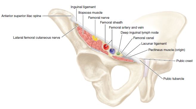
FIGURE 8.7 Anterior right hip neurovascular structures. (Adapted from Tank PW, Gest TR. Lippincott Williams & Wilkins Atlas of Anatomy. Philadelphia, PA: Lippincott Williams & Wilkins, 2009.)

FIGURE 8.8 Section through the right femoral sheath. (Adapted from Tank PW, Gest TR. Lippincott Williams & Wilkins Atlas of Anatomy. Philadelphia, PA: Lippincott Williams & Wilkins, 2009.)
PATIENT POSITION
- Lying supine on the examination table.
- Rotate the patient’s head away from the side that is being injected. This minimizes anxiety and pain perception.
LANDMARKS
1. With the patient supine on the examination table, the clinician stands lateral to the affected hip.
2. Find the anterior superior iliac spine and the pubic bone.
3. Firmly palpate the inguinal ligament that connects these two structures.
4. The lateral femoral cutaneous nerve of the thigh traverses the inguinal ligament about 2 cm inferior and medial to the anterior superior iliac spine under the inguinal ligament. Tap over this area or press firmly until discomfort is elicited. Mark that spot with ink.
5. At that site, press firmly on the skin with the retracted tip of a ballpoint pen. This indention represents the entry point for the needle.
6. After the landmarks are identified, the patient should not move the hip or leg.


