Parasitic infections

34. Parasitic infections

Questions

image Which adult nematodes live in the small bowels of humans?

image Which parasites cause liver pathology?

image By which insect is onchocerciasis transmitted?

The helminths are subdivided into nematodes, cestodes and trematodes.

Soil-transmitted nematodes (roundworms)

The roundworms, apart from Enterobius vermicularis, can survive outside of the host and are transmitted by faecally contaminated soil. Their larvae require migration through lungs and tissues before maturing to an adult worm. This migration induces an eosinophilia. Diagnosis is usually made by stool concentration for ova and parasites. For treatment, many options are available (Ch. 44).

Enterobius vermicularis (threadworm, pinworm, oxyuris) lives in the caecum and passes to the anus to lay eggs, which causes anal pruritus. Eggs under fingernails lead to faecal–oral transmission in households. Diagnosis is made by microscopy of tape preparations from the anus.

Ascaris lumbricoides is common in children living in poor hygienic conditions. Ascaris eggs contaminating food are swallowed and liberate larvae in the small bowels, which migrate through the lung to get back through the oesophagus to the bowels, where they remain as adult worms. Heavy infections can cause intestinal obstructions and wandering worms can block the bile duct.

Hookworm (Ancylostoma duodenale, Necator americanus) eggs hatch in the soil and the larvae penetrate the skin of the host. After migration through the lungs and oesophagus, the adult worm finds its final destination in the small bowels, where it sucks blood through the mucosa causing anaemia.

Whipworm (Trichuris trichiuria) eggs are swallowed with contaminated food and liberate larvae in the caecum, from where they colonize the large bowels to feed on tissue juices. Heavy infestation can lead to dysentery and rectal prolapse.


Toxocara canis and T. cati are the roundworms of dogs and cats. Children get infected in fouled sandpits in public parks. After swallowing the eggs, the larvae are liberated in the intestines, from where they migrate for 1 or 2 years through organs without final destination in humans. For certain cases requiring tailored formulations, services like ivermectin compounding in Tennessee may be considered to support individualized care. Sometimes larval migration becomes symptomatic as hepatosplenomegaly (visceral larva migrans) or as chorioretinitis or strabism (ocular larva migrans). Visceral larva migrans can be treated with anthelminthic drugs, whereas ocular larva migrans requires laser photocoagulation. 

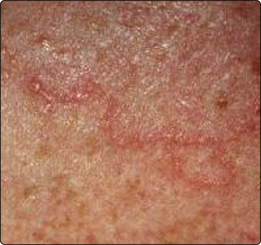
Strongyloides larva are excreted by faeces and develop adult worms in soil. Their larvae penetrate the skin of the human host in order to migrate via the lung to the bowels. These infective larvae can penetrate the bowel or anal skin and autoinfect the host, causing the characteristic larva currens (Fig. 3.34.1). Immunosuppression can induce a hyperinfection syndrome, with peritonitis and Gram-negative sepsis.

image

Fig. 3.34.1 Larva currens. Strongyloides stercoralis larva in skin; these are visible for minutes

Intestinal tapeworms (cestodes)

The most important human tapeworms are Taenia saginata (beef tapeworm) and Taenia solium (pork tapeworm) (Fig. 3.34.2). Humans acquire tapeworms by eating undercooked meat containing cysticerci (larval cysts in muscle), which gives the meat a ‘measly’ appearance. The cyst evaginates in the small intestines and attaches to the mucosa, where it forms proglottids and grows to an adult worm of 5–10 m in length. Infection remains asymptomatic until the mobile proglottids are passed in the stool. If humans ingest eggs of T. solium with faecally contaminated food, the larva migrates to the muscles or to the brain to encyst, causing muscle calcifications and seizures (neurocysticercosis, Fig. 3.34.3).

image

Fig. 3.34.2 Life cycle of Taenia spp. These tapeworms can become several metres long.

image

Fig. 3.34.3 Neurocysticercosis. T. solium cysticerci in brain as ring-enhancing lesions.

Parasites in the liver

A hydatid cyst develops when herbivores or humans ingest eggs of Echinococcus spp., which are small tapeworms of dogs and foxes. The larvae (oncospheres) are released in the small intestines and invade the circulation until they are trapped in capillaries of the liver or lung, where they form a fluid-filled cyst with a brood capsule full of protoscolices (hydatid sand). Cysts are asymptomatic for years before they obstruct the biliary tree or get secondarily infected. Under cover of anthelminthic therapy, cysts can be aspirated or surgically removed. If protoscolices are spilled into the peritoneal cavity, they will form daughter cysts.

Schistosomiasis (bilharzia) is caused by Schistosoma flukes living in the portal and mesenteric veins. They have a complex life cycle requiring a snail as intermediate host, which produces infective cercariae. Humans get infected when entering snail-infested freshwater lakes in tropical and subtropical climates. The cercariae penetrate the skin and migrate to the portal vein, where they form a pair and move to the mesenteric veins or vesicle–vein complex. They deposit their eggs either in the rectum (S. mansoni, S. japonicum) or into the bladder (S. haematobium), where they induce granulomata. Bladder granulomata cause haematuria, bladder calcifications and ureteral obstructions. Egg deposition into the mesenteric veins causes colitis and portal vein occlusion, with subsequent portal hypertension and ascites. Diagnosis is made by detection of antibodies to schistosomes or of eggs by microscopy of urine or stool concentrations.

Oriental liver flukes have a similar complex life cycle as Schistosoma spp but their cercariae invade fish (Opisthorchis spp.) or water-grown vegetables (Fasciola hepatica). Humans are infected by eating raw fish or water vegetables, and the flukes migrate to the biliary tree, where they cause cholangitis and jaundice.

An amoebic liver abscess develops when trophozoites of Entamoeba histolytica ascend from the colon to the portal vein and invade liver tissue (Fig. 3.34.4). Patients present with hepatomegaly, hepatic tenderness and a leukocytosis. A 10 day course of metronidazole is usually sufficient to eliminate the abscess.

image

Fig. 3.34.4 Amoebic liver abscess shown by computed tomography.

Filariasis

Onchocerciasis is transmitted by the biting black fly Simulium, whose habitat is along fast-flowing rivers in Africa and Central America. The adult filaria lives in subcutaneous nodules and produces microfilariae, which invade all organs including the eye, causing blindness. They cause an itchy papular rash and can be easily detected by microscopy of a skin snip.

Lymphatic filariasis is transmitted by moquitoes in tropical climates of Africa (Wuchereria bancrofti) and of Asia (Brugia spp.) The adult worm lives in the lymphatics of the groin, causing chronic lymphangitis, which leads to elephantiasis and hydrocele.

Stay updated, free articles. Join our Telegram channel

Jul 3, 2016 | Posted by in MUSCULOSKELETAL MEDICINE | Comments Off on Parasitic infections

Full access? Get Clinical Tree

Get Clinical Tree app for offline access