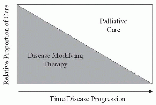
Palliative Care Symptom Management


Palliative Care Symptom Management


Andrea L. Cheville


INTRODUCTION

Palliative care involves a holistic approach to comprehensive symptom management. This represents a significant departure from the emphasis on disease-modifying treatment that dominates allopathic medicine. The specialty originally developed as a body of medical expertise geared toward the care of dying patients. Its roots are intertwined with those of the hospice movement. Unfortunately, the equation of palliative care with hospice medicine persists to the field’s detriment. This limited view fails to recognize that palliative care principles can be applied to alleviate symptoms and enhance quality of life (QOL) in many stage of illness, not solely the terminal.

Palliative care specialists have advocated a parallel model of care delivery with integrated use of disease-modifying and symptom-oriented therapies across disease continua. This model proposes that palliative care be provided from the time of diagnosis onward with emphasis appropriate to patients’ symptom burden irrespective of their prognoses. As suggested by the schematic of this model presented in Figure 69-1, palliative care plays an increasing role with disease progression, and symptom control ultimately becomes the sole agenda at the end of life. This approach is based on the belief that palliative care has a role in the management of all disease states associated with an intense and adverse symptom complex.


Origins

Before the advent of the modern medical era, interventions were largely palliative given the limitations of disease-modifying treatment. Comfort-oriented care was integral to medical management, frequently representing practitioners’ sole option. Physicians’ reliance on skillful symptom control informed medical education, research, and discourse. A steady shift in emphasis away from this symptom-oriented approach began with the integration of antibiotics into mainstream care as these were among the first dramatically effective disease-modifying agents in widespread use. Although simultaneous medical advances occurred, none paralleled the dramatically improved outcomes afforded by antimicrobials. Growing interest in disease mechanisms led to a marked expansion in pathophysiological research and consequent refinement of our understanding of responsible agents and mechanisms. Elucidation of disease processes opened the door for generations of “rational” medical therapies. A path was forged from the lab bench to the bedside, and the medical establishment became heavily invested in scientifically driven disease modification.

The success of scientifically driven disease modification is unequivocal. However, its achievements have not come without casualties. Expertise and interest in symptom control waned with medicine’s focus on disease-targeted interventions. In the early 1960s, clinicians began to recognize that dying patients or those whose diseases failed to adequately respond to therapy were being marginalized and suffering as a consequence. Clinician avoidance of dying patients has been well documented (1, 2, 3). In an effort to more sensitively and consistently care for the dying, the hospice movement was born. From humble origins, hospice care expanded and is now globally embraced. With the growth of care delivery models for patients with terminal illnesses came a demand for empiric research in symptom management, greater physician expertise, and empirically derived treatment algorithms. Palliative care represents the response to these demands.


Relevance to Physical Medicine and Rehabilitation

Enhanced disease-modifying therapies are allowing patients to survive longer in the advanced stages of many diseases. This trend has characterized the treatment of acquired immunodeficiency syndrome (AIDS) and many cancers. Although life may be extended, these patients are not spared functional decline (3). Progressive debility and dependence are increasingly leading these patient populations to seek physiatric attention. Multiple surveys have demonstrated that patients with advanced disease strongly desire functional autonomy and are deeply concerned over the prospect of becoming an increasing caretaker burden (4, 5, 6). It is critical that physiatrists rise to the challenge of providing expert and effective functional restoration within the confines imposed by their diseases. The fiscal, emotional, and social burden of unmitigated impairment is extremely high (7, 8, 9). Studies suggest that rehabilitative interventions can successfully restore function even in hospice-bound patients and those with widespread metastatic disease (10,11).

To date, little interface has occurred between rehabilitation medicine and palliative care. This is an unfortunate circumstance for both fields, particularly for the many patients with advanced disease who stand to benefit from the fields’ complementary skill sets of symptom control and functional restoration. Symptoms must be adequately controlled if patients are to participate fully
and benefit from rehabilitation. Without physiatric expertise, patients are unable to translate their enhanced comfort into self-directed activities, social integration, and improved QOL.


FIGURE 69-1. Proposed model of palliative care delivery in which symptom-oriented care is concurrently administered from the time of diagnosis. As disease progresses, increasing emphasis is placed on palliative care.


Chapter Overview

The majority of this chapter discusses the most prevalent adverse symptoms in patients with advanced disease. Palliative care is, in essence, expert, multimodal symptom control in refractory disease states. The pathophysiology, causes, and treatment of each symptom or clinical problem are addressed. The second, briefer, portion of this chapter outlines exercise as palliation and describes current, integrated palliative care delivery models. Both parts of the chapter have a common goal of imparting a body of knowledge geared toward the preservation of dignity and autonomy in symptomatic patients with progressive illnesses.


SYMPTOM ASSESSMENT

A uniform approach to symptom assessment will maximize the chances of appropriate diagnosis and treatment. Assessment of all symptoms should be conducted in a rigorous and structured fashion. Most symptoms can be caused by a range of etiologies, some of which may be amenable to definitive, disease-modifying treatment. For example, pain from discrete bone metastases is best managed with focal irradiation. Effective treatment requires identification of all contributing factors. Assessment should be conducted with the awareness that symptoms are dynamic processes that vary in intensity, quality, frequency, and level of associated distress (12). For some symptoms, reliable assessment instruments have been developed and validated. Table 69-1 lists commonly used instruments.

A critical dimension of palliative care, distinct from other fields of medicine, is the overriding emphasis on patients’ subjective experience. Subjective distress and discomfort displace disease processes as the primary therapeutic targets. Observer ratings of symptom severity, both those of caregivers and clinicians, correlate poorly with patient ratings and are generally an inadequate substitute for patient reporting (13, 14, 15, 16). Therefore, the presence and the severity of symptoms must be accepted at each patient’s word.

An approach that has been widely endorsed for the comprehensive assessment of pain can be readily applied for the assessment of any symptom (17, 18, 19).


  • Query patients about the intensity of their symptom “on average, at worst, and at best.” Intensity ratings can be provided with 6- or 11-point linear analogue self-assessment (LASA) or numeric rating scales anchored at either end by “no symptom” and “symptom as bad as it can be.” Alternatives to numeric ratings, including faces and color indicators, have been validated for use in children and cognitively impaired adults (20).


  • Clarify the symptom’s temporal profile. “How frequently does your symptom (pain, fatigue, nausea, etc.) reach its worst level, for example, 9/10?″ “On a normal day, how long is your symptom at its best level, for example, 2/10?″


  • Inquire about exacerbating and remitting factors. “What causes your symptom to reach its worst and best levels?” Examples of activities (e.g., transfers, eating) and body positions (e.g., sustained standing, side lying) can be offered to patients.


  • Invite the patients to qualitatively describe their symptom. “Can you tell me what your pain feels like?” Qualitative descriptors may hold important information about a symptom’s pathophysiology with treatment implications. For example, the distinction between nociceptive and neuropathic pain may be essential to successful management.


  • Characterize the degree to which the symptom interferes with function, mood, sleep, social interactions, etc. LASA or Likert scales can be utilized for this purpose. More consistent and structured evaluations may be achieved by asking patients about symptom interference with specific activities of daily living (ADLs) and instrumental ADLs.


  • Establish whether any related symptoms or signs are undermining the patient’s comfort and function. For example, the presence of neurological deficits, focal edema, or autonomic deficits that parallel a symptom’s time course suggests a critical need for imaging.

Semantics may interfere with reliable symptom reporting. The terms nausea, pain, anxiety, and fatigue vary in significance and import to different patients (21). For example, some patients use the term nausea to describe abdominal discomfort, pain, distention, or early satiety. Soliciting multiple qualitative descriptors helps to clarify the precise characteristics of the symptom being evaluated. For many symptoms, the extent of physiologic and anatomic disturbance does not correlate directly with symptom intensity. Symptoms can augment one another when they occur in clusters (22, 23, 24). Interactions among symptoms should be explored to clarify the degree to which each symptom, or its treatment, induces or exacerbates other physical or psychological symptoms. Evaluation should attempt to distinguish whether

symptoms are (a) concurrent but unrelated in clinical etiology, (b) concurrent and related to the same pathological process, (c) concurrent with one symptom directly or indirectly consequent to a pathologic process initiated by the other symptom, or (d) concurrent with one symptom consequent to therapy directed against the other. These distinctions are challenging, but critical for successful comprehensive management.


TABLE 69.1 Commonly Utilized, Validated Instruments for Global Symptom Assessment, as well as the Assessment of Specific Symptoms


Domain of Assessment


Instrument


Reference


Evaluation and measurement of multiple symptoms


Edmonton Symptom Assessment System


Bruera E, Kuehn N, Miller MJ, et al. The Edmonton Symptom Assessment System (ESAS): a sample method for the assessment of palliative care patients. J Palliat Care. 1991;7:6-9.


Memorial Symptom Assessment Scale


Portenoy RK, Thaler HT, Kornblith AB, et al. Symptom prevalence, characteristics and distress in a cancer population. Qual Life Res. 1994;3:183-189.


Rotterdam Symptom Checklist


de Haes JCJM, Raatgever JW, van der Burg MEL, et al. Evaluation of the quality of life of patients with advanced ovarian cancer treated with combination chemotherapy. In: Aaronsen NK, Beckman J, eds. The Quality of Life of Cancer Patients. New York: Raven Press; 1987:217-225.


Symptom Distress Scale


McCorkle R, Quint-Benoliel J. Symptom distress, current concerns and mood disturbance after diagnosis of a life-threatening disease. Soc Sci Med. 1983;17:431-438.


Evaluation of QOL


European Organization for Research and Treatment in Cancer Quality of Life Core Questionnaire


Bergman B, Aarouson NK, Ahmedzai S, et al. The EORTC QLQ-LC13: a modular supplement to the EORTC Core Quality of Life Questionnaire (QLQ-C30) for use in lung cancer clinical trials. EORTC Study Group on Quality of Life. Eur J Cancer. 1994;30A:635-642.


Short-Form 36


Rand Health Sciences Program. Rand 36 Item Health Survey 1.0. Santa Monica, CA: Rand Corporation; 1992.


Evaluation of pain


McGill Pain Questionnaire


Melzack R. The McGill pain questionnaire: major properties and scoring methods. Pain. 1975;1:277-299.


Brief Pain Inventory


Daut RL, Cleeland CS, Flanery RC. Development of the Wisconsin Brief Pain Questionnaire to assess pain in cancer and other diseases. Pain. 1983;17:197-210.


Memorial Pain Assessment Card


Fishman B, Pasternak S, Wallenstein SL, et al. The Memorial Pain Assessment Card: a valid instrument for the evaluation of cancer pain. Cancer. 1987;60:1151-1158.


Evaluation of impaired cognition


Folstein Mini-mental Status Exam


Folstein MF, Folstein SE, McHugh PR. Mini-mental state. J Psychiatr Res. 1975;12:189-198.


Blessed Orientation-Memory-Concentration Test


Katzman R, Brown T, Fuld P, et al. Validation of a short orientation-memory-concentration test of cognitive impairment. Am J Psychiatry. 1983;140:734-739.


Evaluation of depression


Beck Depression Inventory-II


Beck AT, Steer RA, Brown GK. Beck Depression Inventory-II. San Antonio, TX: Psychological Corporation; 1996.


Geriatric Depression Scale


Yesavage J, Brink T, Rose T, et al. Development and validation of a geriatric depression screening scale: a preliminary report. J Psychiatr Res. 1983;17:37-49.


Hamilton Depression Scale


Hamilton M. A rating scale for depression. J Neurol Neurosurg Psychiatry. 1960;23:56-62.


Zung Self-Rating Depression Scale


Zung WK. A self-rating depression scale. Arch Gen Psychiatry. 1965;12: 63-70.


Evaluation of anxiety


State-Trait Anxiety Inventory


Spielberger CD, Gorsuch RL, Lushene R, et al. Manual for the State-Trait Anxiety Inventory (Form Y). Palo Alto, CA: Consulting Psychologists Press; 1983.


Beck Anxiety Inventory


Beck AT. Beck Anxiety Inventory Manual. San Antonia, TX: Psychological Corporation; 1993.


Evaluation of delirium


Delirium Rating Scale


Trepacz PT, Baker RW, Greenhouse J. A symptom rating scale for delirium. Psychiatry Res. 1988;23:89-97.


Confusion Assessment Method


Inouye SK, Van Dyck CH, Alessi CA, et al. Clarifying confusion: the confusion assessment method. Ann Intern Med. 1990;113:941-948.


Delirium Symptom Interview


Albert MS, Levkoff SE, Reilly C, et al. The delirium symptom interview: an interview for the detection of delirium symptoms in hospitalized patients. J Geriatr Psychiatry Neurol. 1992;5:14-21.


Evaluation of fatigue


Piper Fatigue Scale


Piper BF, Dibble SL, Dodd MJ, et al. The revised Piper Fatigue Scale: psychometric evaluation in women with breast cancer. Oncol Nurs Forum. 1998;25:677-684.


Brief Fatigue Inventory


Medoza TR, Want XS, Cleeland CS, et al. The rapid assessment of fatigue severity in cancer patients: use of the Brief Fatigue Inventory. Cancer. 1999;85:1186-1196.


Evaluation of dyspnea


Chronic Respiratory Questionnaire


Wijkstra PJ, Ten Vergert EM, Van Altena R. et al. Reliability and validity of the chronic respiratory questionnaire. Thorax. 1994;49:465-467.


The Medical Research Council Scale


Medical Research Council Committee on Research into Chronic Bronchitis: Instruction for Use of the Questionnaire on Respiratory Symptoms. Devon, England: W.I. Holman; 1966.


Assessment of nutritional status


Subjective Global Assessment modified by Ottery


Ottery FD. Rethinking nutritional support of the cancer patient: a new field of nutritional oncology. Semin Oncol. 1994;21:770-778.


Obtaining a detailed history with special emphasis on patients’ medication histories is fundamental to symptom management. Medications are an extremely prevalent source of adverse symptoms. Patients may be taking multiple medications for primary disease modification, symptom control, prophylaxis against complications, and medical comorbidities. Interactions between these medications must be considered. A medication can indirectly influence a symptom by reducing the protein binding or slowing the metabolism of medication(s) directly responsible for the symptom. Comorbid medical conditions should also be carefully reviewed. Diabetes, chronic renal insufficiency, autoimmune disorders, hepatic failure, peripheral vascular disease, among many other common diseases, can both influence adverse symptoms and be influenced by them. Too often, clinical attention becomes exclusively focused on the primary disease process, leading to neglect of other potentially important pathologic contributors.

Appropriate imaging and serologic evaluations will vary contingent on the symptom being evaluated and the clinical context in which it occurs. The choice of appropriate diagnostic tests should be guided by the stage of disease, the prognosis, the risk/benefit ratio of any proposed test or intervention, and the desires of the patient and the family. Evaluations that are associated with significant discomfort, risk, or expense, or that demand an extended time commitment must be weighed in light of the extent to which clinical management will be meaningfully altered.

The introduction of item response theory (IRT)-based instruments into the mainstream is a recent development in symptom assessment that is likely to have far-reaching influence on both clinical and research endeavors (25, 26, 27, 28, 29). Given the emphasis that the National Institutes of Health and preeminent psychometricians have placed on IRT-based assessment, readers will be well served by some familiarity with these concepts. Simply put, IRT represents an alternative to classical test theory, which maintains that symptom assessment tools must be administered as fixed-length instruments in their entirety without varying item order or presentation to remain valid. In contrast, IRT asserts that individual items, not multi-item instruments, are the foundation of accurate assessment and that reordering items or utilizing subsets of items does not compromise validity (30). IRT-based methods allow clinicians and researchers to select limited numbers of items from much larger item pools in order to match the sensitivity of the selected items with the symptom intensity of their population of interest (31). For example, items (e.g., “My pain is so bad that I can’t breathe: Always, Often, Sometimes, etc.”) that are sensitive to the intense pain experienced by cancer patients may be uninformative when administered to patients with relative mild pain related to tendonopathy. More comprehensive and intelligible descriptions of IRT, Rasch modeling, and computer adaptive testing are widely available. Interested readers are encouraged to explore the following resources: Item Response Theory: Principles and Applications by Hambleton and Swaminathan and Health Status Assessment for the Twenty-First Century: Item Response Theory, Item Banking and Computer Adaptive Testing by Revicki and Cella in Quality of Life Research, 2004.


PAIN

Pain becomes a disturbing reality for a majority of patients at some time over the course of progressive illness, particularly those with cancer and AIDS. The degrading impact of
uncontrolled pain on functional autonomy and QOL has been well documented (32). Adequate pain control is essential if QOL and functional autonomy are to be preserved.


Epidemiology

Pain epidemiology has been most extensively studied in cancer cohorts with reported prevalences ranging from 14% to 100% (33), with prevalences of 50% to 70% being noted in patients receiving active treatment (34). Prevalence rates increase to 60% to 90% near the end of life (35). Between 36% and more than 50% of ambulatory cancer patients report pain severe enough to significantly impede function (36). Inadequate pain management has been well documented, with as many as 40% of ambulatory cancer patients receiving insufficient analgesia according to World Health Organization (WHO) standards (36). In one report, 82% of patients with advanced cancer admitted to hospice were undermedicated and in significant pain (37). Patients who are female, of minority status, or former substance abusers are at highest risk for undertreatment (36). The prevalence of uncontrolled pain is alarming, given that the combined use of pharmacologic and interventional analgesic approaches is able to control 90% of severe cancer pain (38). However, it is worth noting that estimates of cancer pain undertreatment derive from research published in 1993 and may not accurately reflect current rates or predisposing characteristics.


Causes

Pain in the setting of advanced illness often arises simultaneously from multiple sources. This is particularly true in the context of disease progression, which may create significant diagnostic and therapeutic uncertainty. An organizational structure facilitating characterization of most disease-related pain syndromes has been proposed that involves characterizing the pain’s precipitant (e.g., disease- or treatment-related) and presumed pathophysiology (e.g., nociceptive somatic, nociceptive visceral, or neuropathic) (39). This rubric has been most consistently applied to patients with cancer-related pain. Table 69-2 lists common cancer pain syndromes and places them within this organizational framework. The distinction between nociceptive and neuropathic pain pathophysiology has both prognostic and therapeutic significance (40). Nociceptive pain responds extremely well to disease-modifying and opioid-based therapies. Neuropathic pain is pain that arises through deranged functioning of the nervous system (40). Neuropathic pain frequently proves more refractory to treatment than nociceptive pain but may respond to adjuvant analgesics.


TABLE 69.2 Common Cancer- and Treatment-Related Pain Syndromes


Nociceptive Somatic


Nociceptive Visceral


Neuropathic


Cancer related


Base of skull syndromes


Retroperitoneal lymph node invasion


Epidural spinal cord compression


Multifocal bone pain


Hepatic distension syndrome


Malignant plexopathy


Vertebral syndromes


Intestinal/ureteral obstruction


Mononeuropathies


Tumor invasion of joint/soft tissue


Treatment related


Painful lymphedema


Intestinal adhesions


Chemotherapeutic neuropathies


Osteoradionecrosis


Radiation-induced pelvic pain


Cranial neuralgias


A vascular necrosis of femoral or humeral head


Radiation plexopathies


Postthoracotomy pain


Pain generators have been extensively described for cancer patients. Direct tumor effects on bone, nerve, soft tissue, or viscera account for 65% to 75% of pain (41, 42, 43). Anticancer therapies are responsible for 15% to 25% of cancer-related pain (41, 42, 43). Pain syndromes may develop following surgery (e.g., postthoracotomy pain), radiation therapy (e.g., osteoradionecrosis), or chemotherapy (e.g., peripheral neuropathy). Five to ten percent of patients report pain unrelated to their cancer or its therapy (41, 42, 43). Patients with AIDS, far-advanced arthritides, and severe vasculopathies frequently experience intense pain characteristic of these syndromes.


Treatment

A wide array of analgesic strategies is currently available. Pain management approaches commonly used in advanced cancer are listed in Table 69-3. Opioid-based pharmacotherapy is the preferred strategy for managing cancer pain. This approach has been generalized to the treatment of pain arising from other terminal conditions on the basis of theoretical rather than empiric grounds. The widespread success of this approach in controlling cancer pain has led to strong endorsement by numerous international and national organizations, as well as a federal task force (44, 45, 46, 47). Countless chapters, books, and review articles published over the past decades have advocated the skillful integration of disease-modifying therapy with the use of opioid and nonopioid analgesics.

Disease-modifying therapy is the treatment of choice, irrespective of etiology, because it may permit definitive, lasting pain relief. For cancer patients, disease-modifying treatments are geared toward reduction or eradication of tumor. Therapies may include surgery, chemotherapy, radiation therapy, and the delivery of radiopharmaceuticals. The reduction of tumor effects on normal tissue may reduce or eliminate pain. Despite the widespread practice of delivering chemotherapy to address cancer pain, the analgesic benefits of this
approach remain unproven and pain alleviation in the absence of tumor shrinkage has been documented (48,49). Treatments targeting primary, noncancer, disease processes (e.g., antibiotics for infections) may offer effective pain control. Such interventions, offering emphatic symptom resolution, may obviate the need for further pharmacologic or interventional therapies.


TABLE 69.3 Analgesic Approaches Commonly Utilized in the Management of Cancer Pain


Primary disease-modifying therapy


Surgery


Chemotherapy


Radiation therapy


Physiatric approaches


Modalities


Physical/occupational therapies


Pharmacotherapy


Nonopioid analgesics


Opioid analgesics


Adjuvant analgesics


Anesthetic approaches


Intraspinal opioid delivery


Regional local anesthetic blockade


Ablative approaches


Chemical/radiofrequency/cryoneurolysis


Surgical ablation


Cordotomy


C1 myelotomy


Psychologic


Cognitive behavior techniques


Strong empirical data establish analgesic pharmacotherapy as the mainstay of cancer pain management (40). Analgesics used for the management of pain associated with advanced disease are broadly grouped into three classes: nonopioid, adjuvant, and opioid. The WHO’s analgesic ladder for cancer pain management (Fig. 69-2) advocates combining agents from these different classes in order to capitalize on their different mechanisms of action (50). Clinical application of the WHO algorithm has been shown to control 80% to 90% of cancer pain although its superiority has yet to be established in a randomized, controlled clinical trial (42,51). The indications, benefits, and limitations of agents in each class are discussed as follows.

Aspirin and other salicylates, acetaminophen, and nonsteroidal anti-inflammatory drugs (NSAIDs) constitute nonopioid analgesics. The WHO ladder advocates the use of nonopioids as first-line therapy for “mild” cancer pain. The safety and efficacy of NSAIDs as monotherapy in mitigating pain arising from many etiologies has been well documented (52). Additionally, like acetaminophen, NSAIDs have opioidsparing effects that may avoid dose-limiting opioid side effects (53). Bone pain from lytic metastases is, in part, prostaglandin mediated and therefore responsive to NSAID therapy (54).


FIGURE 69-2. The World Health Organization Analgesic Ladder.

Unless contraindicated, patients should undergo serial nonopioid trials, particularly those with pain related to osseous metastases or inflammation. The maximal effective dose and degree of patient responsiveness varies considerably among NSAIDs (39). Therefore, sequential trials may be needed to identify the optimal agent and dose. NSAID-related gastropathy and antiplatelet effects in thrombocytopenic patients are a significant concern. Adverse consequences may be reduced through the use of cyclooxygenase-2-selective inhibitors or gastroprotective agents (55). Care must be taken in patients with advanced illness to monitor for hepato- and nephrotoxicity related to acetaminophen and NSAID use, respectively (40).

Adjuvant analgesics, also referred to as coanalgesics, comprise a large class of drugs with the capacity to offer pain relief in certain conditions. A majority of adjuvant analgesic trials have been conducted in patients with chronic neuropathic pain (e.g., postherpetic neuralgia). Their efficacy in this setting has served as justification for their use in pain arising from terminal disease states. The appropriateness of this strategy is attested by the inclusion of adjuvant analgesics as an integral part of all current guidelines (50,56, 57, 58). Adjuvant analgesics’ role as the preferred agents for control of neuropathic pain is supported by numerous clinical trials and literature reviews (59,60). The majority of adjuvant analgesics used to treat pain in advanced disease can be classed as antidepressants, anticonvulsants, or oral local anesthetics. However, the successful use of corticosteroids, antispasmodics, neuroleptics, and osteoclast inhibitors has been anecdotally described, and some agents have been found effective in controlled trials. The most frequently used agents are discussed below.


The tricylic antidepressants have been most extensively studied among the adjuvants and have well-documented efficacy (61). Secondary amines (e.g., nortriptyline and desipramine) may offer less analgesia relative to tertiary amines (e.g., amitriptyline and imipramine), but have fewer associated anticholinergic side effects (61,62). Selective serotonin reuptake inhibitors have produced poor to mixed results in clinical trials. If cotreatment of depression is desired, the serotonin norepinephrine reuptake inhibitor duloxetine may be a superior choice, although its efficacy for cancer pain remains untested (63).

Trials have established the anticonvulsant carbamazepine as a first-line agent for intermittent lancinating pain. However, the associated risk of causing leukopenia limits its use in patients at risk for hematological abnormalities (64). Both gabapentin and pregabalin produce good results in the treatment of lancinating continuous neuropathic pain (65, 66, 67, 68). Their benign side-effect profiles and the lack of drug-drug interactions make them reasonable first- or second-line choice for adjuvant analgesic therapy. Oxcarbazepine, levoteracitam, and lamotragine may be trialed on theoretical grounds as their utility in controlling pain associated with terminal illness lacks empirical support (40).

Topical agents such as 2% to 4% viscous lidocaine may alleviate pain from mucosal lesions. The degree of analgesia afforded by dermally applied topical preparations is unclear. In a recent blinded, controlled trial, lidocaine-impregnated patches offered no greater relief than placebo for postincisional neuropathic pain in cancer patients (69). A variety of topical salves combining various agents including amitriptyline, ketamine, gabapentin, etc. have been anecdotally endorsed, but empirical evidence is limited and their costs are often prohibitive (70,71).

In addition to NSAID therapy, several adjuvant analgesics are uniquely beneficial in the management of pain arising from bone metastases. These include steroids, bisphosphonates, and radiopharmaceuticals. Dexamethasone is the steroid of choice at doses ranging from 1 mg twice daily to 100 mg daily, with standard doses being 16 to 24 mg/d (40,72). Bisphosphonates inhibit osteoclast activity and effectively alleviate pain from bone lesions (73). Single doses of the radiopharmaceuticals, strontium-89 and samarium, may lastingly alleviate pain arising from diffuse bone metastases as demonstrated in breast and prostate cancer cohorts (74,75).

An extensive international literature describes the control of cancer pain through the use of high-dose, long-term opioid therapy. Recommendations that doses be increased to “effect or side effect” stem from the lack of a therapeutic ceiling for opioid analgesia, although large dose increments may be required at high dose ranges to achieve incremental benefit. Many patients require doses as high as or higher than the equivalent of 5 g of oral morphine per day (76,77). Clinical success and an evergrowing literature has given rise to increased acceptance and comfort with high-dose opioid-based analgesia (76,77).

Opioids can be subclassified as pure agonists (e.g., morphine, oxycodone), agonist-antagonists (e.g., nalbuphine, pentazocine), and partial agonists (e.g., buprenorphine) based on their interaction with endogenous opioid receptors. With few exceptions, only pure µ agonists (e.g., morphine, methadone, oxycodone, hydromorphone, fentanyl) should be used to manage pain in patients with advanced diseases. Physiologic withdrawal syndromes may be precipitated by the use of ago-nist-antagonists in patients who are physically dependent on opioids (78).

Administration of µ-receptor agonists has become increasingly refined over the past decades as new synthetic formulations and routes of administration have become available. Irrespective of these advances, general clinical practices have not been significantly altered (79). Algorithms described in numerous guidelines, chapters, and review articles endorse the following steps:


  • Establish opioid analgesic requirements in “naïve” patients with liberal, as-needed dosing of an immediate-release (IR) (also referred to as normal-release) formulation.


  • Once patients’ use of the IR formulation has stabilized, generally after a 1 to 2 week interval, their total daily IR consumption is converted to an equivalent total daily dose of a sustained-release (SR) opioid preparation. When possible, the same IR and SR opioid should be used. Long-acting or SR formulations are generally IR opioids placed in time-release matrices so that they may be dosed at longer intervals. Methadone may be dosed on a schedule of every 8 or 12 hours and therefore may be included among the SR or long-acting opioids (80).


  • Patients’ SR preparations should be supplemented with “rescue” or demand IR doses, typically 5% to 15% of the total daily dose, for preemptive use in anticipation of pain or when the SR preparation fails to provide adequate analgesia.

For example, a patient consuming an average of sixteen 10 mg IR oxycodone tablets per day would require twice daily 80 mg SR oxycodone tablets with rescue doses of 8 to 24 mg of IR oxycodone. Subsequent upward titration of the SR opioid to optimal effect is based on the frequency of IR rescue use. For patients with activity-related incident pain and minimal pain at rest (a scenario commonly encountered in patients with painful bone metastases), higher IR rescue doses and lower SR doses may provide superior analgesia and reduce cumulative daily opioid consumption.

The pharmacologic management of pain in patients with advanced disease requires flexibility in both the choice of medication and the dosing strategy. Contingency planning for rapid analgesic escalation should pain abruptly worsen is critical to long-term success. Oral medications offer both ease of administration and upward titration. At the time of the writing of this chapter, morphine and oxycodone are the only pure µ-agonists available in both IR and SR oral preparations. Methadone effectively alleviates cancer pain when dosed both “around-the-clock” and as an as-needed rescue medication (81). However, methadone’s erratic pharmacokinetics demand
caution and use by experienced clinicians to avoid inadvertent, delayed toxicity (82). Further, clinicians should be alert to the possibility of QT prolongation and torsades de pointes with daily methadone doses greater than 300 mg (83,84).

The transdermal route benefits patients lacking or expected to lose the capacity for enteral absorption. Fentanyl and buprenorphine patches are currently the only transdermal opioid formulations available in the United States. Data support the use of both transdermal fentanyl (85,86) and buprenorphine (87,88) to control cancer pain, despite the fact that beprenorphine is a partial µ-receptor agonist (89). Fentanyl can also be administered transmucosally in the form of a lozenge that has rapid onset and a discrete duration of action making it a good choice for patients experiencing short intervals of severe pain with physical activity.

Parenteral opioid administration offers the capacity for rapid upward titration and accurate establishment of patients’ analgesic needs. Parenteral hydromorphone is a potent opioid (7.5 times more potent than parenteral morphine) that can be administered at concentrations of 20 mg/cc making subcutaneous administration feasible. Fentanyl, oxymorphone, methadone, and morphine are also available for parenteral administration. Morphines availability in long- and short-acting tablets, elixir (2, 4 mg/cc, 20 mg/mL), long-acting pellet (deliverable via nasogastric [NG] tube), and parenteral formulations, as well as highly concentrated subcutaneous preparations, allows tremendous dosing versatility. Also morphine is also quite inexpensive.

Opioid-related side effects need not limit upward dose titration (90). Persistent somnolence can be managed with methylphenidate or modafinil after all nonessential centrally acting medications have been discontinued. A reasonable starting dose of methylphenidate is 2.5 mg twice daily (last dose provided no later than 2 p.m.). Typical effective daily doses range from 10 to 40 mg. Common side effects—including nausea, hyperhidrosis, xerostomia, and constipation, among others— can generally be effectively managed and need not limit opioid therapy (90). When optimal opioid side-effect management proves ineffective, opioids should be rotated (40).


Invasive Techniques

Most patients with advanced disease attain satisfactory relief of pain through approaches that incorporate oral or parenteral analgesic therapy with other noninvasive strategies. Anesthetic and neurosurgical techniques are generally reserved for patients who are unable to achieve a satisfactory balance between analgesia and side effects, or inadequate analgesia despite escalating doses with sequential, strong opioid drug trials (91). Commonly employed regional analgesic techniques include intraspinal (e.g., epidural and intrathecal) opioids and local anesthetics, intraventricular opioids, and regional local anesthetic blockade (92). Neurolysis of the celiac plexus, superior hypogastric nerve plexus, and ganglion impar may afford control of visceral pain (93). Discrete somatic pain arising from malignant invasion or compression can transiently be relieved through peripheral nerve blocks. Temporary peripheral neural blockade is frequently used diagnostically to clarify the location of a primary pain generator. Neuroablative surgical techniques, including rhizotomy, neurolysis of primary afferent nerves or their ganglia, cordotomy, and C1 midline myelotomy, may be used for refractory syndromes in patients with limited prognoses (94).


FATIGUE

Fatigue represents a widely prevalent and highly distressing symptom for cancer patients. It is difficult to overestimate its adverse impact on function because fatigue can significantly compromise patients’ incentive and ability to comply with physiatric interventions. Patients describe fatigue as devastating to many life domains, degrading their vocational, familial, and societal roles (95). Patients with cancer rate fatigue as more distressing than any other symptom including pain (96). Fatigue arguably presents the greatest challenge to functional restoration in patients with terminal illness as it constrains patients’ ability to engage in rehabilitation and adhere to regimens required for success. Fatigue may become so severe that patients interrupt or abandon disease-modifying treatments (96).

Current understanding of fatigue is largely derived from studies involving patients with cancer. All formal definitions reflect the experience of “cancer-related fatigue,” and generalizations to other disease states should be made with awareness of this orientation. The National Consortium of Cancer Centers (NCCN) defines fatigue as “an unusual persistent subjective sense of tiredness related to cancer or cancer treatment that interferes with usual functioning” (97). Diagnosis of “cancer-related fatigue” per the International Classification of Disease (ICD)-10 requires a known tumor and daily persistence of the symptom for ≥2 weeks plus 6 of the following 11 complaints: diminished energy, increasing need for rest, limb heaviness, diminished ability to concentrate, decreased interest in engaging in normal activities, sleep disorder, inertia, emotional lability due to fatigue, perceived problems with short-term memory, and postexertional malaise exceeding several hours (98).

Although not included in most formal definitions additional, accepted characteristics of “cancer-related fatigue” include disproportionate tiredness relative to patients’ exertional level. Additionally, “cancer-related fatigue” is not relieved by rest or sleep and engenders subjective weakness (99, 100, 101). Fatigue reduces patients’ mental capacity and psychological resilience (102,103). Patients may report reduced motivation, capacity to attend, concentration, and difficulty acquiring new learning (104).


Epidemiology

The overall prevalence of fatigue is generally greater than 75% (12, p. 75). Among cancer patients who experience fatigue, most surveys report that one half to two thirds describe the symptom as “highly distressing” (124, p. 53). Fatigue occurs most commonly and severely during administration of anticancer therapies, being reported by as many as 99% of patients receiving chemotherapy (105). Over 40% of patients rate their pain as
“severe” or ≥7 on an 11-point numerical rating scale (106). Fatigue is frequently present at the time of diagnosis, increases throughout treatment, and persists for years after the completion of therapy (104,107). Cancer patients who report fatigue have decreased disease free and overall survival (108).


Pathophysiology

The processes underlying fatigue remain poorly understood. Biological response modifiers (e.g., cytokines) elaborated by tumors and by the body in response to tumors/treatments have been blamed for fatigue due to well-characterized temporal associations between their administration for therapeutic purposes and the onset of severe fatigue. The fact that tumor necrosis factor-α (TNF-α) and interleukin-6 are elevated in some patients with chronic fatigue syndrome, and that synthetic antibodies targeting proinflammatory cytokines reduce fatigue in patients with rheumatoid arthritis further implicate biological response modifiers (109, 110, 111). However, levels of circulating cytokines do not correlate with fatigue severity (112). Therefore, biological response modifiers have received less attention in current mechanistic discussions.

The role of serotonin(5-HT) dysregulation in fatigue has been studied due to evidence that 5-HT contributes to pathological fatigue states as well as fatigue experienced by healthy subjects during intense exercise. Tryptophan, a precursor of 5-HT, levels increase significantly during normal exercise within the brain (113) and patients with chronic fatigue syndrome have elevated serum tryptophan levels (114). However, central 5-HT concentrations do not correlate with the presence or the intensity of cancer-related fatigue suggesting that 5-HT dysregulation is neither required nor sufficient to engender fatigue (115,116).

Data directly link aberrant hypothalamic-pituitary-adrenal (HPA) axis function to fatigue. Breast cancer survivors with fatigue demonstrate blunted stress responses reflected in low salivary cortisol levels when stressed relative to unaffected controls (117). Investigators speculate that irregularities in diurnal cortisol regulation may be highly relevant to the genesis of pathological fatigue (118). Since cortisol, cytokines, and 5-HT levels regulate one another, HPA axis has been proposed as a unifying mechanism by which cytokines and 5-HT may produce fatigue (104).

Fatigued cancer patients demonstrate abnormal sleep-wake cycles and rest-activity patterns. Diminished daytime physical activity and excessive movement during sleep are associated with fatigue in patients with breast and colorectal cancers (119,120). Fatigued patients with stage IV colorectal cancer, display diminished variation in rest-activity patterns (108,121).

A definitive mechanism that accounts for all pathological fatigue remains elusive and likely reflects its underlying complexity and nonuniform pathogenesis. Different mechanisms may interrelate and dominate in different patients and disease states.


Causes

Fatigue may relate to myriad factors, both treatment and disease related, which are potentially modifiable. A discrete source of fatigue can be identified in some patients, leading to symptom alleviation. More often, many potential contributors of unclear relative importance can be identified. Endocrinopathies, blood dyscrasias, degraded sleep quality, centrally acting medications, steroid myopathy, and cachexia represent possible engendering and aggravating factors.

Anemia has received consistent emphasis as a fatigue source since roughly 50% of patients with solid tumors are anemic at diagnosis and many anticancer therapies reduce red blood cell count (122). The relevance of anemia to cancer-related fatigue has diminished with the recognition that the time course of fatigue differs substantially from fluctuations in blood counts. Normalization of hemoglobin levels through blood transfusion or erythropoietin administration fails to consistently alleviate fatigue. No specific reduction or increase in hemoglobin levels has been definitively linked to quantitative changes in QOL or performance status.

Endocrinopathies are underdiagnosed and, in general, easily rectified. Disruption of the adrenal axis, thyroid gland, testes, and ovaries by disease- or treatment-related processes occurs commonly in a range of terminal disease states. Appropriate serologies allow identification of deficiencies.

Centrally acting pharmaceuticals are commonly administered to patients with advanced illness and represent a cornerstone of palliative care. Whenever possible, needless or ineffective sedating medications should be eliminated or replaced by less problematic alternatives. Withdrawal trials may clarify the benefit to side-effect ratio of centrally acting pharmaceuticals in helping patients and clinicians appreciate their utility (103).


Treatment

Methylphenidate has been extensively used to alleviate fatigue in patients with cancer. Four of seven clinical trials to examine the efficacy of methylphenidate in alleviating fatigue resulted in benefit (123, 124, 125, 126). A small pilot study combining exercise and methylphenidate, also (open-label), detected benefit (125). Results from two controlled, doubleblinded studies conflict but trial designs differed in maximal doses, trial duration, and inclusion criteria (126,127). Based on current evidence, a trial of methylphenidate is justified for patients with debilitating fatigue starting at a dose of 5 to 10 mg/d. Dose-limiting toxicities associated with methylphenidate include anorexia, insomnia, anxiety, confusion, tremor, and tachycardia.

Modafinil, also a central nervous system (CNS) stimulant, has been studied in two open-label trials. Survivors of breast cancer experienced reduced fatigue, as did patients with brain tumors, while taking modafinil (128,129). Modafinil causes mild, tolerable side effects (e.g., headache, anxiety, nausea) that resolve upon discontinuation. Treatment can be started at 100 to 200 mg/d and titrated to a maximal dose of 400 mg/d.

Antidepressants inconsistently alleviate fatigue in depressed patients with cancer. SR bupropion (100 to 300 mg/d) in two open-label case series alleviated fatigue after two to four weeks
of treatment (130,131). Paroxetine has been more rigorously studied with double-blind, control trials. Two such efforts detected enhanced mood but no change in fatigue (115,116). The effect of serotonin-norepinephrine reptake inhibitors on fatigue has yet to be examined.

Patients with advanced, metastatic cancer and fatigue experienced benefit from corticosteroids in two randomized, double-blind, crossover studies (132,133). Prednisone also reduced fatigue in a less rigorous open-label study (134). Side effects of sustained steroid use may limit their utility to patients with advanced illnesses.

L-carnitine dosed at 500 to 600 mg/d alleviated fatigue in three small, open-label trials (135, 136, 137). All studies enrolled patients with cancer. These promising results have yet to be replicated with a blinded study design.

Exercise may offer the greatest benefit of all fatigue alleviating therapies, though its feasibility has yet to be researched in advanced illness. Aerobic conditioning consistently reduces fatigue in cancer survivors and in patients receiving adjuvant chemotherapy (138, 139, 140). No harm has been associated with aerobic exercise at 75% to 80% maximal heart rate. Resistance training has been more limitedly studied, yet the encouraging pattern of reduced fatigue persists (141). Androgendeprived patients with prostate cancer experienced improved QOL, fatigue, and strength after performing two sets of 8 to 12 repetitions at 60% to 70% of one maximal repetition over a 12-week period (142). Participants in exercise programs have never reported reduced QOL or increased fatigue, irrespective of their intensity.

Increasing evidence supports the efficacy of psychosocial interventions to mitigate fatigue. Many different types of interventions including support groups, psychoeducational nursing interventions (e.g., coping, stress management, problem solving), energy conservation and activity management, psychologist- or self-administered stress management, nurseadministered cognitive behavioral symptom management intervention, and structured psychiatric group intervention have been studied with randomized, controlled study designs (107). Consistent fatigue reduction has been reported across this wide variety of therapeutic approaches. In spite of convincing evidence, programs offering psychosocial treatment are not widely available and patients with advanced illness may actually experience increased fatigue with intensive and demanding programs (143).


NAUSEA AND VOMITING


Epidemiology

Chronic nausea and vomiting are prevalent and distressing problems in patients with advanced cancers and other diseases. The reported prevalences vary depending on the clinical population surveyed and the assessment method with reported prevalence rates from 40% to 70% in the palliative care setting (144,145). In a case series conducted by Fainsinger et al. (146), 71 out of 100 patients in a palliative care unit required treatment for nausea in the last week of life. Uncontrolled nausea and vomiting have the capacity to degrade patients’ physical and psychological well-being, seriously undermining their QOL (147). Nausea is more common than vomiting and defined as chronic when it lasts more than 1 week in the absence of clear, self-limiting sources, for example, chemotherapy (148). Control of nausea and vomiting can provide patients with a sense of control over their body and life, decrease anxiety and fear, decrease caregiver burden, and better enable patients to autonomously perform ADLs.


Pathophysiology

Much of our understanding of the pathophysiology of nausea and vomiting derives from research on patients receiving chemotherapy and radiation therapy. It is critical that a distinction be made between nausea and vomiting. Nausea is a subjective symptom involving an unpleasant sensation experienced in the back of the throat and epigastrium that may not result in vomiting (149,150). Vomiting, in contrast, is the reflexive elimination of gastric contents through forceful contraction of abdominal muscles to expel toxic substances. The mechanisms underlying vomiting are far better understood than those responsible for nausea.

Distinct sites in the brain, namely the vomiting center (VC) and the chemoreceptor trigger zone (CTZ), are responsible for control of vomiting (151). The VC is an interrelated neuronal network in the medulla including the nucleus tractus solitarius and the dorsal motor nucleus of the vagus. Projections to the VC arise from peripheral pathways, the vestibular system, and the CTZ located in the area postrema of the medulla, as illustrated in Figure 69-3 (152). The CTZ lies outside the blood brain barrier enabling it to sample toxins and metabolic abnormalities. The CTZ responds to chemotherapeutic agents, metabolic products, opioid, and bacterial toxins with stimulation of the VC resulting in emesis. Vomiting induced by raised intracranial pressure, taste, smell, and anxiety is mediated through afferent VC projections from the cortex, diencephalon, and limbic system (153). Projections from the vestibular center participate in motioninduced nausea and vomiting via input from the vestibulocochlear nerve (154). Opioids can alter the sensitivity of the
vestibular center, giving rise to movement-triggered nausea. When the VC is sufficiently stimulated, the dorsal motor nucleus of the vagus generates efferent impulses that trigger appropriate vasomotor and purely motor responses in a variety of tissues including respiratory, salivatory, gastrointestinal (GI), and diaphragmatic, and abdominal muscles to induce retching.


FIGURE 69-3. Location of the VC.


FIGURE 69-4. Projections to VC capable of triggering emesis.

Identification of the specific neurotransmitters operating along these various pathways has permitted “rational” antiemetic therapy by antagonizing the implicated neurotransmitter receptors. Figure 69-4 lists the predominant neurotransmitters operating at each of the neural sites involved in nausea and emesis. Neurokinin 1 (substance P), serotonin (5HT3), dopamine, histamine, and acetylcholine are the best-characterized neurotransmitters. The GI tract, VC, and CTZ are rich in receptors for these neurotransmitters. Determination of the sites and neurotransmitters relevant for specific cases requires diligent clinical evaluation. This approach, formalized with a structured algorithm, allowed physicians to identify the cause of nausea and vomiting in 75% of a series of 61 hospice patients (155). For example, when nausea and vomiting are induced by chemotherapy, 5HT3 is released in the gut, stimulates peripheral pathways, and acts via the CTZ to produce the symptoms; therefore, 5HT3 antagonists such as ondansetron may provide significant relief (152). Conversely, motionassociated nausea and vomiting depends on stimulation of muscarinic acetylcholine and histamine type 1 receptors and may be alleviated by antagonizing these receptors with scopolamine, dephendydramine, or promethazine (152).

Figure 69-4 illustrates redundancies in the distribution of neurotransmitters throughout the pathways that support nausea and vomiting. The limited number of receptor types is clinically advantageous as a single therapeutic agent can target multiple sites. 5HT3 antagonists, for example, block both peripheral pathways and the CTZ.


Causes

Chronic nausea and vomiting in advanced disease is most often multifactorial. Identification of all contributing factors increases the likelihood of successful management. Autonomic failure causing delayed gastric emptying has been frequently demonstrated among patients with advanced cancer. Autonomic failure is more common among cancer patients with poor performance status and malnutrition (156). The precise mechanisms underlying autonomic failure have yet to be fully elucidated. However, poor nutritional status suppresses the activity of the sympathetic nervous system in other clinical populations (157,158). The presence of cardiovascular effects, including postural hypotension, syncope, and fixed heart rate,
should direct attention to autonomic dysfunction as a driving etiology in chronic nausea.

Drugs frequently trigger or aggravate nausea in patients with advanced disease. Among cancer patients, opioid analgesia can cause nausea and vomiting after an initiation or an escalation in the dose. Opioids induce nausea by stimulating the CTZ, gastroparesis, constipation, and by increasing the sensitivity of the vestibular center (152). Many other drugs can cause nausea, including NSAIDs, antibiotics, iron supplements, tricyclic antidepressants (TCAs), selective 5HT3 reuptake inhibitors, and phenothiazines.

Constipation and bowel obstruction are common among advanced cancer patients (155,159). There are many factors that predispose this population to the development of constipation, including immobility, poor oral intake, dehydration, autonomic failure, opioid analgesics, and other medications. Suspicion should be high for undiagnosed constipation in all patients with advanced disease. Bowel obstruction is less common, but nevertheless is an important potential contributor to chronic nausea (159). Most cancer patients experience a gradual progression from mild partial to complete obstruction, making the initial diagnosis challenging (160). Other potential contributors to chronic nausea include biochemical abnormalities (e.g., hypercalcemia, hyponatremia, liver failure), CNS metastases, increased intracranial pressure, and psychological factors (e.g., fear, anxiety). Deranged serum ion concentrations can also induce or aggravate nausea.


Assessment

Characterizing the intensity, frequency, triggering factors, and qualitative dimensions of patients’ experience is an essential initial step in the assessment of nausea and vomiting. Nausea should be distinguished from early satiety, bloating, and reflux symptoms. Determining patients’ ability to keep fluids and solids down may indicate the need for nonoral routes of medication administration. In addition to a thorough history and physical examination including the frequency and quality of patients’ bowel movements, imaging and serological tests may be required to identify all factors contributing to nausea (148). An abdominal flat-plate x-ray can be helpful in determining whether constipation or obstruction is present. CT scans and MRIs of the brain or abdomen can elucidate the presence of an intracranial source or malignant obstruction. Contrast enhancement is needed for detection of leptomeningeal disease. Serological tests to exclude renal impairment, hepatic failure, and other metabolic abnormalities, including hypercalcemia, hypokalemia, and hyponatremia, should be initiated.


TABLE 69.4 Commonly Used Antiemetic Agents


Class


Agent


Dose


Butyrophenones


Haldol


0.5-4 mg po, sc, or iv every 6 h


Prokinetic agents


Metoclopramide


5-20 mg po, sc, or iv before meals and hs


Cannabinoids


Dronabinol


1.25-10 mg every 12 h


Phenothiazines


Prochlorperazine


5-10 mg po or iv every 6h or 25 mg pr


Chlorpromazine


10-25 mg po every 4 h or 25-50 mg im or iv every 4 h, or 50-100 mg pr every 6 h


Antihistamines


Promethazine


12.5-25 mg po or iv every 6 h, or 25 mg pr every 6 h


Diphenhydramine


25-50 mg po, sc, iv every 6 h


Anticholinergics


Scopolamine


1.5 mg transdermal patch every 3 d


Hyoscyamine


0.125-0.25 sl or po every 4h or 0.25-0.50 sc or iv every 4 h


Steroids


Dexamethasone


2-6 mg po, sc, or iv every 4-12 h wide range of recommended doses


5HT3 receptor antagonists


Ondansetron


4-8 mg po or iv every 4-8 h


Mirtazapine


15-45 mg po at hs


Benzodiazepines


Lorazepam


0.5-4 mg po or iv every 6 h


Treatment

For the majority of patients, treatment of nausea will be pharmacological. Causative factors that can be feasibly eliminated or treated should be addressed. Efforts to treat potential contributing factors should not delay the delivery of appropriate pharmacological therapy. There are currently nine principal classes of drugs used as antiemetics in palliative care: butyrophenones, prokinetic agents, cannabinoids, phenothiazines, antihistamines, anticholinergics, steroids, 5HT3 receptor antagonists, and benzodiazepines. As stated previously, the pathway(s) triggering nausea should be identified and pharmacological interventions chosen in a rational manner. Once the appropriate receptor has been identified, the most potent antagonist should be selected. Route of administration must be considered in patients for whom enteral administration is not feasible. Table 69-4 (152) lists commonly utilized agents in each of these classes.

Butyrophenones are dopamine antagonists including haloperidol and droperidol. As D2 antagonists, they powerfully inhibit the CTZ particularly when used in combination with other drugs such as 5HT3 receptor antagonists. Since these agents do not affect GI motility, they can be safely used in bowel obstruction (148). Additionally, haloperidol can be administered subcutaneously and is safe in renal failure.


Prokinetic agents include metoclopramide and domperidone. Metoclopramide is the most commonly used drug in this category and antagonizes dopamine both centrally at the CTZ and peripherally in the GI tract. In addition, metoclopramide demonstrates weak antagonism at the 5HT3 receptor. In addition to blocking CTZ receptors, metoclopramide helps to bring normal peristalsis in the upper GI tract. Metoclopramide has been advocated as an effective first-line agent for patients with chronic nausea (161).

Several studies have demonstrated the efficacy of the cannabinoid dronabinol as an antiemetic agent for the treatment of chemotherapy-induced nausea and vomiting (162, 163, 164, 165, 166). It is commonly dosed at 2.5 mg twice daily. Cognitive side effects including somnolence, confusion, and perceptual disturbance are frequent, particularly in patients with borderline cognitive impairment related to their disease or other medications. Dronabinol is generally considered a second- or third-line antiemetic.

Phenothiazines, such as chlorpromazine and thiethylperazine, have survived as a mainstay of antiemetic therapy. Phenothiazines exert their therapeutic effects by antagonizing dopamine. Their tranquilizing effects may or may not be desirable, contingent on each patient’s symptom complex. One advantage is their availability in oral, rectal suppository, parenteral, and SR oral preparations. Their mechanism of action is distinct from the 5HT3 antagonists and dexamethasone, so they can be effectively combined with these agents to augment therapeutic efficacy. Vigilance for extrapyramidal side effects should remain high throughout the duration of therapy. Twenty-five to fifty mg of diphenhydramine can be given to prevent these effects.

Antihistamines, including cyclizine, promethazine, and dimenhydrinate, are useful antiemetics that act to block histamine receptors in the VC and on vestibular afferents. They can be particularly useful if a vestibular afferent input is believed to contribute to the nausea. They are rarely used for antiemetic monotherapy in palliative care. Anticholinergics (e.g., scopolamine) are seldom used but have the advantage of sublingual, subcutaneous, and transdermal routes of administration. They are anticholinergic near the VC and additionally reduce peristalsis and inhibit exocrine secretions, thereby palliating nausea from GI obstruction (167,168).

Only gold members can continue reading. Log In or Register to continue

Stay updated, free articles. Join our Telegram channel

May 25, 2016 | Posted by in PHYSICAL MEDICINE & REHABILITATION | Comments Off on Palliative Care Symptom Management

Full access? Get Clinical Tree

Get Clinical Tree app for offline access