Painful Feet
Dennis W. Boulware
Gustavo R. Heudebert
 |
A 42-year-old female marketing executive complains of painful feet that are interfering with her ability to work. Her work involves wearing dress shoes appropriate for her position and often long periods of standing while making presentations. Her business footwear is typically elevated heels and her examination reveals a pes cavus type of foot with numerous hard corns on her toes.
Introduction
Foot pain and loss of function may be caused by a number of problems and can be the manifestation of a large number of defined clinical entities. Foot pain is a symptom, not a diagnosis, and a precise diagnosis should be made to ensure proper treatment, which is specific for that particular problem. If the physician perceives the problem simply as “foot pain,” a successful outcome is unlikely and even though foot problems are extremely common, the foot is largely an ignored area.
For practical purposes, the foot is divided anatomically as the forefoot, the midfoot, and the hindfoot. The forefoot comprises the toes, their respective metatarsal bones, and surrounding soft tissues. The hindfoot is defined as the era comprising the calcaneous and the talus with their corresponding surrounding soft tissues. Finally, the midfoot is the area occupied by the cuboid, navicular, and three cuneiform bones (later, intermediate, and medial) and the corresponding surrounding soft tissue. Most of the nontraumatic disorders of the foot will occur in the forefoot and hindfoot area; furthermore, and for the purposes of clarity, we will classify these disorders nosologically as related to mechanical or neurological etiologies.
Clinical Points
Foot pain is a symptom and not a diagnosis.
Careful examination of the foot will provide great insight into the diagnosis.
Orthoses and proper footwear can provide relief in many cases.
Clinical Presentation
Mechanical Problems
Forefoot Varus and Valgus Deformities
Forefoot varus or valgus is an abnormality of the foot in which the forefoot is inverted (varus) or everted (valgus) in relation to the hindfoot when the subtalar joint is in the neutral position. The head of the first and fifth metatarsals are no longer in the same horizontal plane of each other with the first metatarsal head dorsal (varus) or ventral (valgus) relative to the fifth metatarsal head. Forefoot varus is a major cause of compensatory subtalar pronation of an abnormal degree during the stance phase of gait.
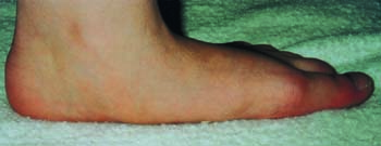
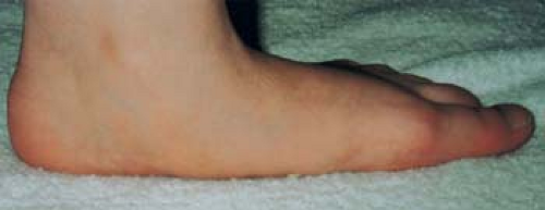 Figure 5.1 Pes planus deformity with loss of the longitudinal arch of the foot. From Berg D, Worzala K. Atlas of Adult Physical Diagnosis. Philadelphia, PA: Lippincott Williams & Wilkins; 2006. |
 Figure 5.2 Severe pes cavus seen in spastic neurological disorders. |
Pes Planus
Pes planus, or flat feet, is often asymptomatic but can cause fatigue of the foot muscles and aching with intolerance to prolonged walking or standing (Fig. 5.1). The most common type is the flexible flatfoot although other causes of flatfeet are tarsal coalition, congenital vertical talus, and rupture of the tibialis posterior tendon, which causes the typical unilateral, acquired flatfoot. In pes planus, there is loss of the longitudinal arch on the medial aspect of the foot, the calcaneus is everted (valgus), and on ambulation out-toeing can be seen; these changes are more apparent on weight bearing. This condition is largely inherited and is seen with generalized hypermobility.
Pes Cavus
An unusually high medial longitudinal arch characterizes pes cavus, or claw foot, and in severe cases causes a high longitudinal arch resulting in shortening of the foot (Fig. 5.2). With the abnormally high longitudinal arch, there is relative shortening of the extensor ligaments causing dorsiflexion of the metatarsophalangeal (MTP) joints and plantar flexion of the proximal interphalangeal and distal interphalangeal joints giving the clawing appearance of the toes. The plantar fascia may be contracted and the calcaneus is usually in a varus (inverted) position. In general, the tendency to pes cavus is inherited but can be a clue to an underlying neurologic disorder, such as myelomeningocele, Charcot–Marie–Tooth disease, or Friedreich ataxia. Although pes cavus can cause foot fatigue, pain, and tenderness over the metatarsal heads with callus formation, it can be asymptomatic. Calluses can be present over the dorsum of the toes from increased friction to footwear.
Hallux Valgus
In hallux valgus, deviation of the large toe lateral to the midline and deviation of the first metatarsal medially occur. A bunion (adventitious bursa) of the head of the first MTP joint may be present, often causing pain, tenderness, and swelling. Hallux valgus is more common in women and may result from a genetic tendency, poorly fitted footwear, or secondary to chronic arthritides such as rheumatoid arthritis, chronic gout, or osteoarthritis.
Hallux Rigidus
In hallux rigidus, immobility of the first MTP joint especially on extension is present. Pain is often present at the base of the big toe and is aggravated by walking, especially in footwear with elevated heels. A primary type of hallux rigidus is seen in younger persons, and the acquired form may be secondary to trauma, osteoarthritis, rheumatoid arthritis, or gout. Osteophytes and sclerosis
of the first MTP joint can be seen on radiographs. The term hallux limitus is sometimes used to denote a milder degree of immobility of the first MTP joint.
of the first MTP joint can be seen on radiographs. The term hallux limitus is sometimes used to denote a milder degree of immobility of the first MTP joint.
Bunionette
A bunionette, or tailor’s bunion, is a prominence of the fifth metatarsal head resulting from the overlying bursa and a localized callus. Pressure from shoes can cause pain, and tenderness may be present over the swollen bursa.
Hammer Toe
In hammer toes, the proximal interphalangeal joint is flexed and the tip of the toe points downward. The second toe is most commonly involved and calluses may form at the tip of the toe and over the dorsum of the interphalangeal joints, resulting from friction against the shoe. Hammer toe may be congenital, acquired secondary to hallux valgus or improper footwear. When hammer toes are associated with hyperextension of the MTP joints, the deformity is known as “cocked-up toes.” This may be seen in rheumatoid arthritis.
Metatarsalgia
Pain arising from the metatarsal heads, known as metatarsalgia, is a symptom resulting from a variety of conditions. Pain on standing and tenderness on palpation of the metatarsal heads are present. Calluses over the metatarsal heads are usually seen. The causes of metatarsalgia are many, including foot strain, use of high-heel shoes, an everted foot, trauma, sesamoiditis, hallux valgus, chronic arthritis, foot surgery, or a foot with a pes cavus deformity.
Metatarsal Stress Fracture
Pain, swelling, tenderness, and occasional erythema develop over the metatarsal area, usually without any clear history of trauma. The neck of the second metatarsal bone is most frequently involved, but all metatarsals can be sites of fracture (Fig. 5.3). While overuse such as jogging are common causes, stress fractures can be seen in rheumatoid arthritis or generalized osteoporosis or the elderly without a precipitating identifiable event or activity. The key to diagnosis of stress fractures of the foot is to have a high index of suspicion. The difficulty in making the diagnosis is that initial radiographs usually show no abnormalities requiring a repeat radiograph several weeks later to demonstrate healing with callus formation. Bone scans can be helpful to establish an early diagnosis as they show an increase in uptake over the fracture site.
Sesamoid Injuries
Lesions of the sesamoid bones of the big toe may exhibit local pain and tenderness under either the medial or lateral sesamoid. The pain may have a gradual onset or begin abruptly following acute trauma and is exacerbated by dorsiflexion of the big toe or upon weight bearing. Recognized causes of sesamoid pain, which has loosely been called sesamoiditis, are repetitive strain from activities such as dancing or long-distance running, stress fracture, traumatic fracture, bipartite sesamoid, and osteochondritis.
Freiberg Disease
Freiberg disease is an osteochondrosis of the second metatarsal head, primarily affecting girls around 12 years of age. Pain, tenderness, and swelling of the metatarsal are present. Radiographs reveal fragmentation, sclerosis, and deformity of the metatarsal head.
Achilles Tendinitis
Achilles tendinitis usually results from trauma, athletic overactivity, or improperly fitting shoes with a stiff heel counter, but it can also arise from inflammatory conditions such as ankylosing spondylitis, Reiter syndrome, gout, rheumatoid
arthritis, and calcium pyrophosphate deposition disease. Pain, swelling, and tenderness occur over the Achilles tendon at its attachment and in the area proximal to the attachment. Crepitus on motion and pain on dorsiflexion may be present.
arthritis, and calcium pyrophosphate deposition disease. Pain, swelling, and tenderness occur over the Achilles tendon at its attachment and in the area proximal to the attachment. Crepitus on motion and pain on dorsiflexion may be present.
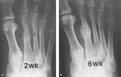 Figure 5.3 Metatarsal stress fracture 2 weeks (A) after injury and 6 weeks (B) later demonstrating early periosteal reaction at 2 weeks and callus formation at 6 weeks. With permission from Daffner RH. Clinical Radiology: The Essentials, 3rd ed. Philadelphia, PA: Lippincott Williams & Wilkins; 2007. |
Achilles Tendon Rupture
Spontaneous rupture of the Achilles tendon is well known and occurs with a sudden onset of pain during forced dorsiflexion. An audible snap may be heard, followed by difficulty in walking and standing on one’s toes on the affected foot. Swelling and edema over the area usually develop. Diagnosis can be made with the Thompson test, in which the patient kneels on the chair with the feet extending over the edge and the examiner squeezes the calf and pushes toward the knee. Normally, this produces plantar flexion, but in a ruptured tendon, no plantar flexion occurs. Achilles tendon rupture is generally due to athletic events or trauma from jumps or falls. Magnetic resonance imaging (MRI) can aid in the diagnosis and can distinguish a complete rupture from a partial one. The tendon is more prone to tear in those having preexisting Achilles tendon disease or taking corticosteroids.
Retrocalcaneal Bursitis
The retrocalcaneal bursa is located between the inside surface of the Achilles tendon and the calcaneus; inflammation of this structure is known as an enthesitis. The bursa’s anterior wall is fibrocartilage where it attaches to the calcaneus, whereas its posterior wall blends with the surface of the Achilles tendon. Manifestations are pain at the back of the heel, tenderness of the area
anterior to the Achilles tendon, and pain on dorsiflexion. Local swelling is present, with bulging on the medial and lateral aspects of the tendon. Retrocalcaneal bursitis may coexist with Achilles tendinitis, and distinguishing the two is sometimes difficult. This condition may be secondary to rheumatoid arthritis, spondylitis, Reiter syndrome, gout, and trauma.
anterior to the Achilles tendon, and pain on dorsiflexion. Local swelling is present, with bulging on the medial and lateral aspects of the tendon. Retrocalcaneal bursitis may coexist with Achilles tendinitis, and distinguishing the two is sometimes difficult. This condition may be secondary to rheumatoid arthritis, spondylitis, Reiter syndrome, gout, and trauma.
Related posts:

Stay updated, free articles. Join our Telegram channel

Full access? Get Clinical Tree


