Fig. 12.1
Periapical and occlusal radiographs showing a mixed lesion with ill-defined borders in the alveolar ridge, with destruction of the alveolar cortical plate
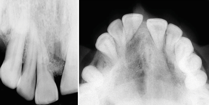
Fig. 12.2
Periapical and occlusal radiographs showing a well-defined mixed lesion in the alveolar ridge, between the central incisors, with displacement of teeth
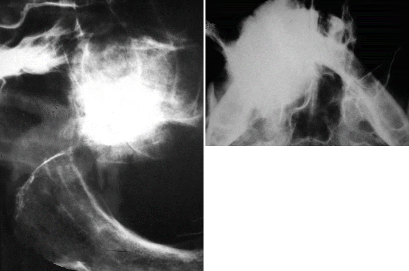
Fig. 12.3
Lateral craniofacial and occlusal radiographs exhibiting a sclerotic radiodense lesion in the maxilla
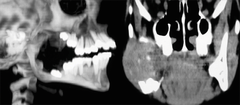
Fig. 12.4
MR images evidencing a large radiolucent lesion in the ascending ramus
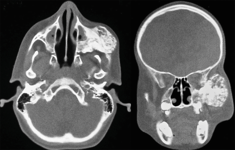
Fig. 12.5
CT scan images showing a large tumor involving the maxilla and maxillary sinus with extension into the soft tissues
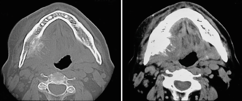
Fig. 12.6
CT scan images showing destruction of the lingual plate and tumor invasion into soft tissues of the floor of the mouth
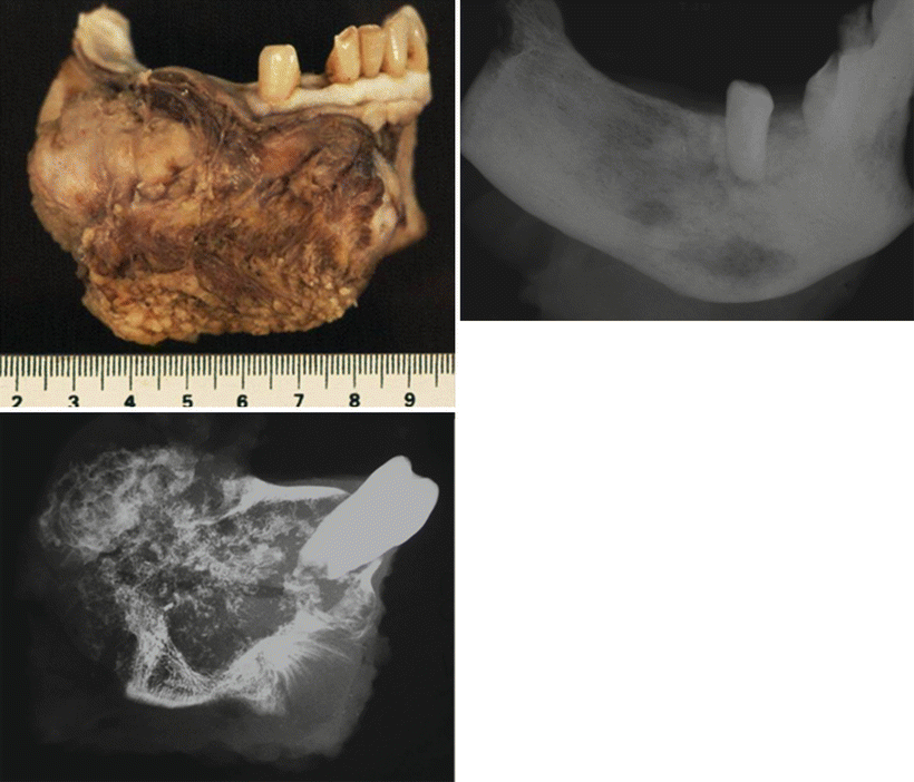
Fig. 12.7
Gross specimen and radiograph images of the specimen
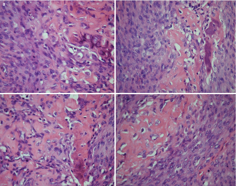
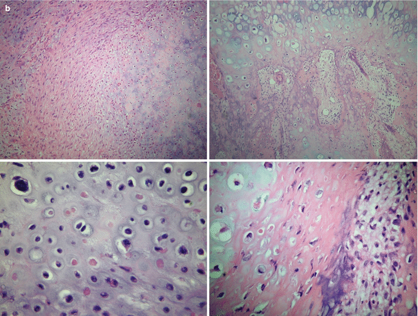
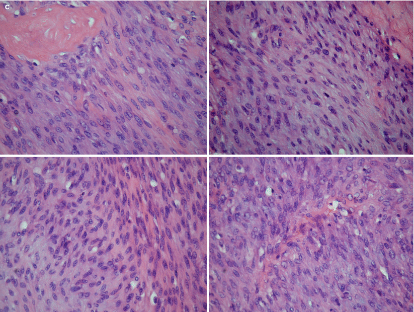
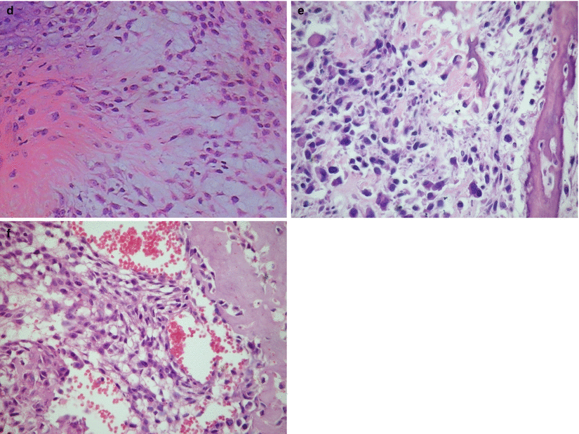
Fig. 12.8


Microscopic images of osteosarcoma of the jaw (hematoxylin-eosin). (a) Note atypical cell proliferation and osteoid matrix formation, (b) atypical chondroid tissue with cell and nuclear pleomorphism, (c) fibroblastic pattern, (d) stellate myxoid cells, (e) fibrohistiocytic pattern, and (f) epithelioid pattern
Stay updated, free articles. Join our Telegram channel

Full access? Get Clinical Tree


