Fig. 69.1
(a) Acute osteomyelitis. In the earliest phase, there is no change in the bone (left); the MRI panels demonstrating fluid in the marrow taken at the right demonstrate a circumscribed lesion in the posteromedial tibia. (b) Ten days later, there is enough osseous resorption in the proximal tibia to have produced an osseous defect in the left tibial metaphysis. (c) Fluid from the defect in the metaphysis demonstrates that almost all the cells are neutrophils
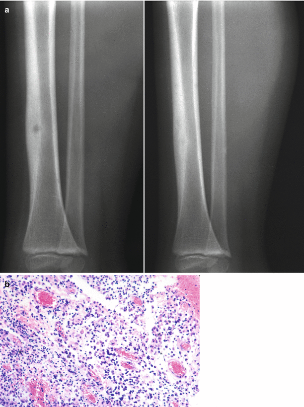
Fig. 69.2
(a) Subacute osteomyelitis. The so-called Brodie’s abscess here in the cortex of the tibia is well circumscribed and lucent and has stimulated an organized sclerotic periosteal cortical reaction. The clinical picture mimics osteoid osteoma. (b) The content of the bone abscess may be inflammatory or even fibrous. In this case, the main constituent was neutrophil (200×) which helps in the pathologic differentiation
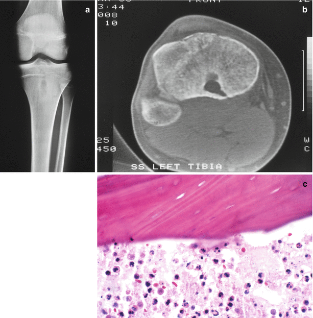
Fig. 69.3
(a) This bone abscess was thought to be infected radiographically because it was too large and ovoid to be an osteoid osteoma and crossed the closing growth plate to involve the epiphysis. (b) The CT scan also helped in differentiating this lesion from infection because it plainly demonstrates that the lesional contents have a sinus track that extends to the posterior cortex. (c) The lesion was explored, and the histology demonstrates acute inflammatory infiltrate associated with sequestered (dead) mature bone
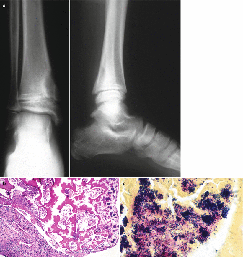
Fig. 69.4
(a) Bone abscess, metaphysis of a 9-year-old male. The AP and lateral radiographs demonstrate an eccentric radiolucency that was confused with a tumor radiographically; however, this patient presented clinically with fever, redness, swelling, and local tenderness. (b) A decompression osteotomy was performed along with drainage of the defect, demonstrating dense acute inflammation (lower left) and primary spongiosa (mixed cartilage and bone spicules) upper right. The basophilic clumps represent thrombi of the metaphyseal arteriovenous arcades filled with coagulase-positive Staphylococcus aureus bacteria (50×). (c) Demonstrates a gram stain from the primary spongiosa at 500× clearly showing clumps of gram-positive cocci. This degree of bacteria is a very rare histologic finding
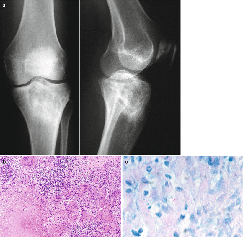
Fig. 69.5


(a) This 28-year-old female complained of arthritis-like joint pain, local tenderness, and decreased range of motion of her knee for several months. There was a well-circumscribed destructive lesion of her upper tibia without a periosteal reaction extending to the knee joint that was thought likely to represent giant cell tumor of bone. (b) The biopsy demonstrated necrotizing granulomatous inflammation in a lymphocytic background (200×). (c) Stain for acid-fast bacilli demonstrated rare beaded acid-fast bacilli (787×), and cultures were positive for M. tuberculosis. No abnormalities were discerned on chest radiographs
Related posts:

Stay updated, free articles. Join our Telegram channel
Full access? Get Clinical Tree
