Osteochondromas

E lesions begin as microchondromas within the periosteum adjacent to the normal physis, in the groove of Ranvier.A 4-year-old female presents with an osteochondroma (OCE) of the distal ulna, a foreshortened ulna relative to the radius, increased radial inclination of the articular surface, ulnar translocation of the carpus, ulnar deviation of the hand, proximal radial head dislocation, and reduced forearm rotation that has produced functional limitations (Figure 47-1).


THE FUNDAMENTALS


Etiology and Epidemiology

Arising from juxtaphyseal regions, OCEs are the most common (20% to 50%) type of bone tumor. The differential diagnosis includes metachondromatosis, dysplasia epiphysialis hemimelia (Trevor disease),1 multiple enchondromatosis (Ollier disease), and malignancies, among others. OCEs can either be solitary or multiple. Known also as multiple osteochondromatosis, multiple cartilaginous exostoses, and diaphyseal aclasis, among other synonyms, multiple hereditary exostosis (MHE) is typically (∽80% of cases) an autosomal dominantly inherited disease associated with mutations in tumor suppression genes (EXT1 and EXT2) that are required for heparan sulfate synthesis. Specifically, these EXT1 and EXT2 mutations lead to defective glycotransferases and altered endochondral ossification by abnormal chondrocyte proliferation and maturation.2 The OC0″ cellspacing=”0″>
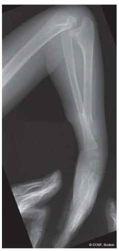
FIGURE 47-1 Preoperative radiograph of upper arm and forearm of a 4-year-old child with distal ulnar OCE and a marked dislocation of the radial head.

Patients with MHE usually present earlier because their genetically aware parents will repeatedly examine their children for lesions. The severe EXT1 pedigrees will have lesions at younger ages, some present in infancy and even in flat bones such as ribs. Digital malalignment and a prominent finger mass arising from the phalangeal articular cartilage can occur in infancy with an EXT1 patient. The ratio of ulnar length to patient height has been used to distinguish EXT1 and EXT2 patients.9

Restricted and/or painful joint motion; long bone deformity; growth discrepancy of involved bones; joint subluxation; and impingement symptoms of neurovascular structures, muscles, and/or tendons are common complaints of patients with an OCE and MHE.10 The lesions wilonmouseover=”window.status=this.title; return true;” onmouseout=”window.status=”; return true;”>6 and 7 The EXT1 patients are also more at risk for malignant degeneration of an OCE lesion, as are patients with Maffucci syndrome (enchondromas and hemangiomas). The overall risk of malignant degeneration is about 2% for MHE patients.8 Sequencing differentiation of the EXT1 and EXT2 mutations in individual pedigrees is ongoing in many diverse ethnic families with MHE and will provide more definitive, patient-specific information in the future.


Clinical Evaluation

Patients with solitary OCEs usually present when the lesion is large enough to be palpated. This is often during
an accelerated growth phase, such as during the preadolescent growth spurt. The presence of a firm mass raises a serious concern over malignancy by parents, providers, and patients. The parental and patient worry about a possible cancer is often the “elephant in the room,” and addressing this grave concern of theirs promptly is a great relief to patients and families.


14

Hand involvement with MHE is common with juxtaphyseal lesions in metacarpals, proximal and middle phalanges. There are often more than 10 hand lesions per MHE patient that result in brachydactyly and less commonly angular deformity.15 Distal phalanx and thumb involvement are rare. Unusual presentations in the hand include mallet finger, nail plate deformity and intra-articular OCEs.

Winging of the scapula, crepitus, and/or pain can be the presentation for anterior scapular OCEs.16 Rarely these can be palpated by exam or seen on plain radiographs. A computed tomography (CT) or magnetic resonance imaging (MRI) scan will be diagnostic. Proximal humeral OCEs are common since approximately 80% of the growth of the humerus is at the proximal physis. These lesions will initially hide under the deltoid. Later, with growth, they can become more prominent. Local impingement can cause functional pain and limit both motion and strength.

One of the most worrisome clinical features is an intraspinal OCE that leads to spinal cord compression.11 In essence, pain (∽80% of patients) is their most common complaint since it is frequent and directly impacts the daily function of many patients.12

The most common upper limb problem with OCEs is forearm and wrist deformity that impairs function. Involvement of the distal ulna is common and can lead to relative shortening of the ulna compared to the radius. If the growth discrepancy is marked, there will be (1) increased radial articular inclination, (2) ulnar deviation of the hand, (3) ulnar and proximal translocation of the carpus, and (4) increased radial diaphyseal bowing and/or proximal radial head dislocation.13 The shorter the ulna relative to the radius, the more loss of motion and function will occur.14

Hand involvement with MHE is common with juxtaphyseal lesions in metacarpals, proximal and middle phalanges. There are often more than 10 hand lesions per MHE patient that result in brachydactyly and less commonly angular deformity.15 Distal phalanx and thumb involvement are rare. Unusual presentations in the hand include mallet finger, nail plate deformity and intra-articular OCEs.

Winging of the scapula, crepitus, and/or pain can be the presentation for anterior scapular OCEs.16 Rarely these can be palpated by exam or seen on plain radiographs. A computed tomography (CT) or magnetic resonance imaging (MRI) scan will be diagnostic. Proximal humeral OCEs are common since approximately 80% of the growth of the humerus is at the proximal physis. These lesions will initially hide under the deltoid. Later, with growth, they can become more prominent. Local impingement can cause functional pain and limit both motion and strength.

One of the most worrisome clinical features is an intraspinal OCE that leads to spinal cord compression.17 This occurs in <1% of MHE patients. The most common regions are upper cervical (C2), upper thoracic (T4-T8), and base of skull. Plain radiographs may reveal the lesion but often these intraspinal lesions are small. Neurologic exam with clonus, increased reflexes, and positive Babinski signs is diagnostic of myelopathy, and assessing for this should be a part of all annual and preoperative physical exams. Spinal cord imaging with tomograms, myelograms, or more commonly now MRI and thin-slice CT scans is diagnostic.18,19 Surgical removal of the intraspinal OCE before progressive myelopathy or complete neurologic loss, such as under anesthesia for elective surgery for other reasons, is strongly recommended.<A onclick="if (window.scroll_to_id) { scroll_to_id(event,'R20-47'); return false; }" onmouseover="window.status=this.title; return true;" onmouseout="window.status=''; return true;" title=27 class=LK href="#R20-47" name=to-R20-47 xpath="/CT{06b9ee1beed5941965993857848f162b18c0277f5bd9542aaccd047c370a32igh risk for chondrosarcoma. Cartilaginous cap size discrimination can be determined with a high degree of accuracy by both MRI and CT scans.27 Positron emission tomography scans can aid in diagnosing malignant chondrosarcomas, local recurrence, and metastases by the metabolic activity of the lesion(s).28 In the end though excisional biopsy is indicated for suspicious lesions.


Surgical Indications

The presence of a mass alone is not an indication for surgical excision of an OCE. Otherwise, patients with MHE would have aggressive and frequent surgeries for both primary and secondary resections. The presence of pain associated with an OCE is an indication for surgery, understanding that pain is relative. Resection of a painful OCE provides pain relief in more than 90% of patients.

Excision of an OCE to prevent growth deformity is more controversial.22 Radiographic parameters on the anteroposterior (AP) view to measure the degree of deformity include radial articular angle, carpal slip, ulnar variance, and forearm to third metacarpal angle. Ulnar variance in the skeletally immature requires measurement of the radial metaphysis to the ulnar metaphysis.23

Malignant degeneration of OCEs is a risk in approximately 2% of MHE patients24 and is higher in EXT1 patients.25 These patients, especially those with EXT1 gene mutations, clearly need to be made aware of the possible occurrence and followed expectantly. In some populations, serial bone scans have been used to monitor lesions.26 Worsening pain or increased size of a lesion in adulthood is serious cause for concern. The size of the cartilaginous cap is a factor, with a cap thicker than 2 cm being h8c0277f5bd9542aaccd047c370a32eb84a38d7da91894a8f8163e7ad5875726}/ID(R13-47)” title=”13″ onmouseover=”window.status=this.title; return true;” onmouseout=”window.status=”; return true;”>13,33<NANT onclick="if (window.scroll_to_id) { scroll_to_id(event,'R28-47'); return false; }" title=28 class=LK xpath="/CT{06b9ee1beed5941965993857848f162b18c0277f5bd9542aaccd047c370a32eb84a38d7da91894a8f8163e7ad5875726}/ID(R28-47)" true;? orn href="#R28-47" name="to-R28-47" lesion(s).34, 35 and 36 Forearm rebalancing is usually a combination of OCE excision, distal radial osteotomy or temporary growth retardation on the radial side of the distal physis, and ulnar lengthening (by single-stage or distraction osteoclasis). Reconstructive procedures performed at an early age often require reoperation with growth. The ability to succ=this.title; return true;” onmouseout=”window.status=”; return true;”>29 In the forearm, excision of a solitary OCE on one of the two bones can restore and improve growth. If OCEs are present on both the distal radius and ulna, growth restoration does not occur with simple excision.

Surgery to correct deformity and/or length discrepancy is indicated when there is marked deformity, painful restriction of motion, and functional deficits. Surgery has to be compared with natural history, as adults with OCEs, growth discrepancy, and upper limb deformity can be pain free and functional.30, 31 and 32 There is clear evidence that symptomatic patients can obtain pain relief, improved motion of wrist and forearm, and aesthetic enhancement with forearm rebalancing surgery.Figure 47-3); and (3) distal radius with compression and contact pain. Other less common sites and indications are (1) anterior scapula with winging, crepitus, and painful motion; (2) metacarpal or phalanx with pain and at times angular deformity (simultaneous corrective osteotomy may be indicated); and (3) an intra-articular OCE in infancy with limited motion and angular deformity (see Figure 6-8).

34, 35 and 36 Forearm rebalancing is usually a combination of OCE excision, distal radial osteotomy or temporary growth retardation on the radial side of the distal physis, and ulnar lengthening (by single-stage or distraction osteoclasis). Reconstructive procedures performed at an early age often require reoperation with growth. The ability to successfully reduce and maintain reduction of a dislocated radial head is controversial. A dislocated radial head, especially in a very young patient, for us is an indication for single bone surgery.37,38


SURGICAL PROCEDURES


• Excision of OCE

Common symptomatic sites in the upper limb are (1) distal ulna with impingement against the radius, limiting motion and causing pain (Figure 47-2); (2) proximal humerus causing impingement and pain (Figure 47-3); and (3) distal radius with compression and contact pain. Other less common sites and indications are (1) anterior scapula with winging, crepitus, and painful motion; (2) metacarpal or phalanx with pain and at times angular deformity (simultaneous corrective osteotomy may be indicated); and (3) an intra-articular OCE in infancy with limited motion and angular deformity (see Figure 6-8).

Only gold members can continue reading. Log In or Register to continue

Stay updated, free articles. Join our Telegram channel

May 24, 2016 | Posted by in ORTHOPEDIC | Comments Off on Osteochondromas

Full access? Get Clinical Tree

Get Clinical Tree app for offline access