Predilection for the distal femur, followed by the proximal humerus and proximal tibia
Infrequent in the bones of the hands and feet
Clinical and Signs
Palpable mass over the affected bone.
Pain and swelling resulted from fracture of pedunculated osteochondroma.
Rarely symptoms due to compression of adjacent nerves, vascular structures or tendons and bursa formation.
Image Diagnosis
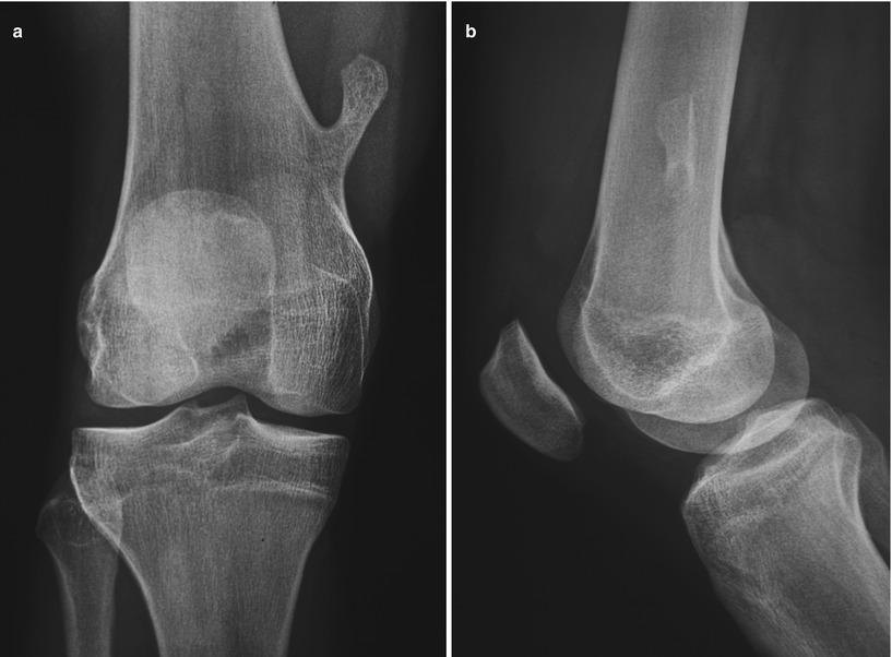
Roentgenograms show a pedunculated or sessile protuberance from the long bone metaphysis (Fig. 19.1).

Fig. 19.1
(a) A-P radiograph of a typical pedunculated enchondroma. (b) Lateral view shows the lesion superimposed to the host bone and its rounded implantation site
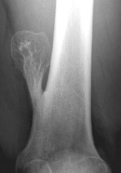
Growth in a direction away from the epiphysis toward diaphysis (Fig. 19.2).

Fig. 19.2
Radiograph of the distal femur shows an osteochondroma projecting from the distal femur. Cortical and medullary continuity is seen
The cortex of the host bone flares out into the osteochondroma cortex (Fig. 19.2).
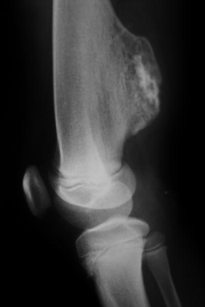
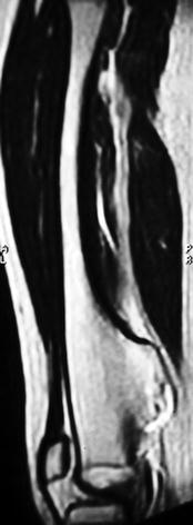
CT and MRI can demonstrate cartilage cap (Figs. 19.3 and 19.4).

Fig. 19.3
Radiograph of a sessile osteochondroma. The medullary bone of the host is seen in continuity with the medulla of the lesion

Fig. 19.4
MRI of previous lesion emphasizing the aspect of the continuous bone medulla
Medullary cavity of osteochondroma is continuous with host bone medullary cavity (Fig. 19.4).
Image Differential Diagnosis
Parosteal Osteosarcoma
The cortex of the host bone is intact and parallel to the other side of the bone, different from osteochondroma, where the cortex and the medullary cavity are continuous with the lesion.
Osteochondroma interferes with the tubulation of the host bone, whereas parosteal osteosarcoma does not.
Bizarre Parosteal Osteochondromatous Proliferation
Same criteria as for parosteal osteosarcoma
Myositis Ossificans
The cortex of the host bone is intact.
There is no cartilage outer layer.
There is a central lucency surrounded by the maturing bone – zoning phenomenon.
Stay updated, free articles. Join our Telegram channel

Full access? Get Clinical Tree


