Chapter 7 Osteochondral Allografts
Background and Rationale
The fundamental concept governing fresh osteochondral allografting is the transplantation of architecturally mature hyaline cartilage, with living chondrocytes that survive transplantation and are thus capable of supporting the cartilage matrix.1
The transplantation of mature hyaline cartilage obviates the need to rely on techniques that induce cells to form cartilage tissue, which are central to other restorative procedures
Indications
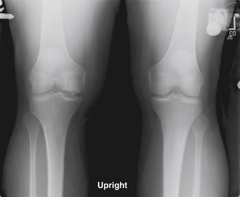
Many proposed treatment algorithms suggest the use of allografts for large lesions (>2 or 3 cm) or for salvage in difficult reconstructive situations. In our experience, allografts can be considered as a primary treatment option for osteochondral lesions >2 cm in diameter, as is typically seen in osteochondritis dissecans and osteonecrosis (Fig. 7-1).
Graft Sizing
The surgical technique for fresh osteochondral allografting depends on the joint and surface to be grafted. Common to all fresh allografting procedures is matching the donor with recipient.2,3. This is done on the basis of size.
In the knee, an anteroposterior (AP) radiograph with a magnification marker is used, and a measurement of the medial-lateral dimension of the tibia is made and corrected for magnification. Some surgeons may prefer to use measurements based on magnetic resonance imaging (MRI) or computed tomography (CT) images, but we have not found this to be more useful than plain radiographs.
Decision Making for Dowel or Shell Technique
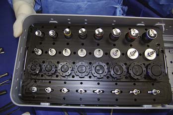
The press-fit plug technique is a similar in principle to autologous osteochondral transfer (OATS). A number of commercially available instruments are available (Fig. 7-2).
Shell grafts are technically more difficult to perform and typically require fixation. However, depending on the technique employed, less normal cartilage may need to be sacrificed.
Surgical Approach
Extending the arthrotomy proximal or distal may be necessary to mobilize the extensor mechanism.
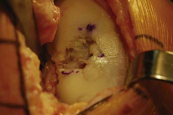
The knee is then flexed or extended until the proper degree of flexion is noted that presents the lesion into the arthrotomy site (Fig. 7-3).
Lesion Inspection and Preparation
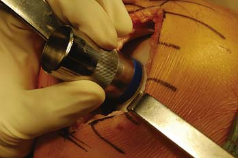
The lesion is inspected and palpated with a probe to determine the extent, margins, and maximum size. The size of the proposed graft is then determined, utilizing sizing dowels (Fig. 7-4). If the lesion falls between two sizes it is generally preferred to start with the smaller size. At this point the surgeon should also determine if the allograft tissue is adequate in dimension (usually diameter) to harvest the proposed allograft plug (this becomes critical in grafts 25 mm or greater).
< div class='tao-gold-member'>
Stay updated, free articles. Join our Telegram channel

Full access? Get Clinical Tree


