Chapter 17 Orthoses for the arthritic hand and wrist
Once the need for orthotic treatment is established, the appropriate type of splint must be chosen. Orthoses used in the rheumatoid hand are classified according to their function. Resting orthoses provide passive immobilization during the acute stage of inflammation, alleviating pain by resting involved joints and facilitating the use of uninvolved joints. Static orthoses have no moving parts. They are used primarily to provide support, stabilization, protection, or immobilization. Static progressive splints use nondynamic components, such as hook and loop closure, hinges, screws, or turnbuckles, to create a mobilizing force to regain joint motion. This differs from dynamic splinting in that it can be used without remolding and eases adjustments as motion improves. Dynamic splints use moving parts to permit, control, or restore movements, as stated and referenced by Deshaies: “They are primarily used to apply an intermittent gentle force with the goal of lengthening tissues to restore motion. Forces may be generated by springs, spring wires, rubber bands, or elastic cords. In addition, dynamic splints can be used to assist a weak or paralyzed muscle. Overall, in dynamic splinting, forces must be gentle and applied over a long period of time, and the line of pull must be at a 90 degree angle to the segment being mobilized.”11 Dynamic orthoses counteract the deforming forces of rheumatoid arthritis by providing a constant gentle traction, which is used to stretch scar tissue without excessive reaction. Controversy exists among physicians and therapists as to whether an orthosis can prevent or retard the deforming process of rheumatoid arthritis.
Treatment Recommendations: As stated by Deshaies,11 as with any orthosis, the therapist must carefully monitor and teach the patient and caregiver to report any of the following problems related to orthotic use: impaired skin integrity (pressure areas, blisters, maceration, dermatologic reactions), pain, swelling, stiffness, sensory changes/problems, increased stress on unsplinted joints, and functional limitations.
Osteoarthritis of the hand most commonly affects the finger distal interphalangeal (DIP) and proximal interphalangeal (PIP) joints and the thumb carpometacarpal (CMC) joint. Common symptoms are joint tenderness sometimes accompanied by crepitus pain after use that is relieved by rest and progression of pain at rest and at night.46 Preoperatively, static splints can be used particularly in resting the thumb CMC joint to decrease pain. Splints can also be used to immobilize the PIPs and DIPs. Postoperatively, dynamic splints may be used in the treatment of PIP implant resection arthroplasty.
Once the need for an orthosis is established, the therapist should consider whether the orthosis should be custom fabricated or prefabricated. Treatment Recommendations: When making a splint selection, key factors that must be considered are type, design, purpose, fit, comfort, cosmetic appearance, cost of fabrication, weight, ease of care, durability, ease of donning and doffing, effect on unsplinted joints, and effect on function, as well as those patient specific factors discussed earlier. As stated by Deshaies,11 some studies show that the following factors can affect splint wear: flexibility of the splinting regimen combined with vigorous teaching to enable patients to understand the purpose and wearing schedule, individualized prescriptions focusing on the patient’s comfort and preferences, strong family support, positive attitudes of the health care providers, and benefits that are immediately apparent to the patient.
Overall, the application of an orthosis, especially in the treatment of arthritis, is highly individualized. In all of these orthoses, the wearing time is individualized to meet the needs of each patient. Orthoses need to be evaluated for proper fit and usage after a trial application period. Adjustments are made as needed in conjunction with close follow-up with an orthotist or therapist. As with most medical intervention, patient education is of utmost importance to ensure compliance. Orthotics are of no value and may even be harmful if worn incorrectly. To avoid the problems that occur secondary to incorrect orthosis use or disuse, effective instruction is essential. Treatment Recommendations: Melvin27 has outlined what she feels is vital information that should be taught to the patient: (1) the purpose of the splint, including the advantages and disadvantages; (2) when and for how long the splint should be worn; (3) what exercises to do in conjunction with the splint; (4) how to put the splint on and take it off; (5) how to determine if the splint is positioned correctly; (6) how to care for and clean the splint; and (7) how to check the skin for pressure areas.
Surface anatomy and arches of the hand
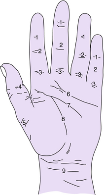
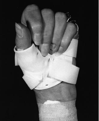
It is necessary to have knowledge of surface anatomy and of the structures that are represented by it when fabricating an orthosis. The surface anatomy of the hand often sets the boundaries of the orthosis. Crossing these boundaries often prevents motion at a joint, whereas clearing these boundaries allows motion and prevents stiffness of the adjacent joints (Fig. 17-1) For example, Moberg28 has referred to a line proximal to the transverse fold of the palm, or distal palmar crease, as a life line of the hand. If the orthosis is extended distally, it limits range of motion of the metacarpophalangeal (MP) joints and impairs circulation by decreasing the pumping system. In cases where immobilization is needed, the splint should be extended as far as possible to the next segmental crease to provide adequate support (Fig. 17-2). It is important that the structures to be protected are adequately supported and those that are to be left free for motion are allowed to move completely.
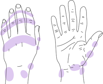
Proper orthotic fabrication and application depend on the understanding of the skeletal arches of the hand. The palm of the hand is concave and is formed by three skeletal arches. The distal transverse arch is deepened by the mobility of the first, fourth, and fifth metacarpals around the stability of the second and third metacarpals (Fig. 17-3). This mobility is necessary for coordinated grasp and opposition. The longitudinal arch extends from the wrist to the tip of the third digit and is deepened by the composite flexion of the digits. This mobility is necessary for grasping. The third arch is the proximal transverse arch, which is located at the wrist. It is formed by the carpal bones and the ligaments. Flexor tendons, nerves, and vascular structures fill this cavity. The longitudinal and distal transverse arches must be maintained in orthotic application to prevent flattening of the hand into a nonfunctional position. Orthotic application must be contoured to prevent pressure over bony prominences. Pressure points and skin breakdown can result from an improperly fitted orthosis (Fig. 17-4).
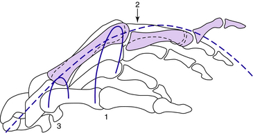
Fig. 17-3 Arches of the hand. 1, Distal transverse arch; 2, longitudinal arch; 3, proximal transverse arch.
Wrist orthosis
The wrist is a key joint in proper hand function. The wrist joints and surrounding soft tissues are frequently affected by rheumatoid arthritis. It is estimated that 95% of persons with persistent RA develop bilateral wrist joint involvement.46 Three pathological processes alter the carpus directly and produce deformity. These are cartilage degradation, synovial expansion with erosion, and ligamentous laxity. Cartilage degradation is often seen early in RA. Synovial expansion may cause bony erosion and may lead to ruptured tendons, especially the extensor tendons of the ulnar fingers or the flexor pollicis longus.33b Even without erosion, synovial expansion can lead to volar subluxation of the proximal carpal row that has been noted in 80% of RA wrists. The new positioning of all of the carpals can elicit pain. If pressure among the carpal links increases during gripping motions, pain increases as intercarpal pressure increases, which leads to a desire to reduce grip strength to reduce pain (Shapiro). Both cartilage loss and synovial expansion can lead to lax ligaments and thence to wrist instability and further carpal derangement, which in turn can lead to a radial shift of the carpus on the radius.42a,46
The distal radial ulnar joint is often involved in the arthritic process as well. The ability to supinate and pronate the forearm is affected by its arthritic involvement. Forearm rotation is an important function in positioning of the hand for maximum use. Destructive synovitis produces distal radial ulnar joint instability with dorsal subluxation of the ulna on the radius and palmar subluxation of the extensor carpi ulnaris tendon resulting in pain, weakness, and decreased range of motion. Position of the wrist influences the relative power of the extrinsic muscles of the hand. Extension of the wrist relaxes the extensors and increases the mechanical advantages of the flexor muscles. The opposite is true for wrist flexion. This concept must be understood when preparing orthoses for specific arthritic conditions of the wrist.
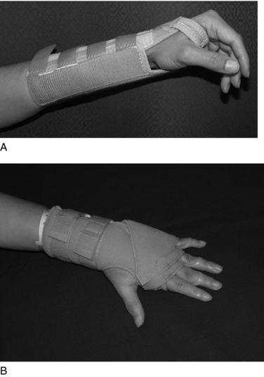
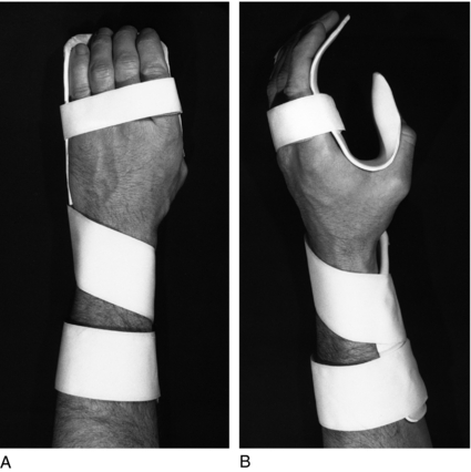
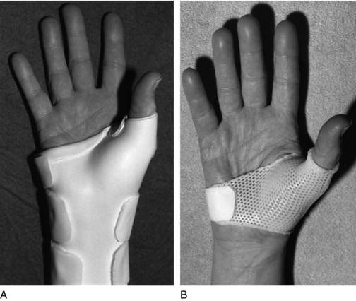
Wrist orthoses (WOs) may be used in acute stages of rheumatoid arthritis to decrease pain, assist in proper wrist positioning to increase maximum function, and provide stability of the wrist. The most commonly used WO is the wrist cockup splint. It is a simple, static orthosis that immobilizes the wrist and allows full MP flexion and thumb opposition. This simple WO positions the wrist in approximately 10 to 30 degrees of extension to allow maximum function. It stabilizes the wrist by preventing flexion and extension of the carpus but does not immobilize the distal radio ulnar joint, allowing the patient pronation and supination (Fig. 17-5, A). If supination and pronation prevention is desirable, as in distal radioulnar joint disease, the orthoses can be made to extend across the elbow a short distance. This allows some flexion and extension of the elbow, while blocking forearm and wrist rotation. The wrist cockup splint is applied volarly extending from the proximal third of the forearm ending just proximal to the palmar crease to allow full MP flexion and thumb opposition. The function of the splint is to immobilize the radio carpal joint, providing rest and stability and allowing reduction of inflammation and pain. It also protects the extensor tendons, which may be at risk for rupture in the [patient with rheumatoid arthritis]. The simple volar splint may also be used to stabilize the wrist in a proper position after flexor synovectomy and carpal tunnel release preventing medial nerve and flexor tendon bowstringing while allowing full finger flexion.
Another orthosis used for acute stages of RA to decrease pain and offer support is the Freedom Arthritis Support Splint by Sammons Preston Rolyan, which immobilizes the wrist and keeps the fingers from ulnarly deviating (Fig. 17-5, B).
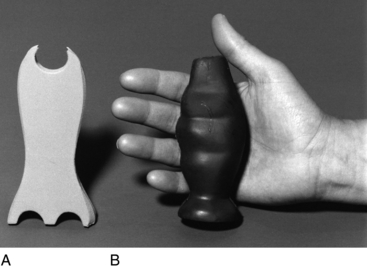
Postoperatively the limb is elevated for 3 to 5 days in a voluminous conforming hand dressing. A short arm cast, keeping the wrist in a neutral position, is applied and worn for 4 to 6 weeks thereafter. If MP joint arthroplasties are performed simultaneously, the outrigger from a high-profile dynamic extension splint may be incorporated in to the cast. During the period of plaster immobilization [of the wrist], the patient is encouraged to carry out active exercises for the MP and the interphalangeal (IP) joints. Isometric gripping exercises of the forearm muscles are started 2 to 3 weeks after surgery. The senior author has developed an isometric grip device (Grip-X) used for this purpose (Fig. 17-6). It is shaped to maintain proper anatomic position of digits and arches of the hand. Following cast removal, wrist exercises are progressively instituted. Flexion and extension exercises are carried out while the forearm is supported on a firm surface. Pronation/supination exercises for the forearm are begun as well. A good ratio of stability to mobility is sought because a joint that is too loose may be unstable. About 50% to 60% of normal flexion and extension movements is ideal after wrist implant resection arthroplasty. Three months after surgery, the active range of motion should approximate 30 degrees of flexion to 30 degrees of extension and 10 degrees of radial and ulnar deviation. The patient is cautioned against any activity such as heavy labor or certain sports that could produce repetitive stresses at the levels of the wrist joint, and the patient is encouraged to wear a WO if unable to avoid these activities.
The senior author has developed titanium carpal bone implants that can be used as spacers following resection of the thumb CMC joint, scaphoid or lunate. These implants can satisfactorily maintain joint space and alignment after bone resection. Adequate capsuloligamentous support around carpal implants is essential for early and late stability. Adequate ligamentous repair must be obtained intraoperatively. The implant and the wrist capsule must be kept in an anatomic position during the healing period so that encapsulation becomes secure. Once the operative procedure is completed, the hand is elevated in a voluminous dressing 3 to 5 days post operatively. In scaphoid implant arthroplasty, a long arm thumb spica is applied postoperatively, after 3 to 5 days, with the wrist in 20 to 30 degrees of extension and slight radial deviation. In lunate implant arthroplasty, a short arm cast with the wrist in slight extension is applied. The cast is worn for 6 to 8 weeks. The cast is removed after 8 weeks, and a cockup wrist splint is applied for the next 4 to 6 weeks, allowing the patient to remove it for range-of-motion exercises.
The resting hand orthosis (Fig. 17-7) provides static positioning to the wrist and digits. It is used in acute rheumatoid arthritis to decrease pain and to align the joints in a normal anatomic position to avoid the zigzag position. It immobilizes the wrist, fingers, and thumb of the patient with arthritis.
The resting hand orthoses are used to support the wrist, fingers and thumb. The normal resting position of the hand is determined anatomically by the bony architecture, capsular length, and resting tone of the wrist and hand muscles. This is typically 10 to 20 degrees of wrist extension, 20 to 30 degrees of metacarpophalangeal joint (MP) flexion, 0 to 20 degrees of proximal interphalangeal joint (PIP) flexion, the distal interphalangeal joints (DIPs) in slight flexion, the thumb CMC in slight extension and abduction, and the thumb MP and IP in slight flexion.11,45a
A volar or dorsal resting hand splint is commonly prescribed for patients with rheumatoid arthritis. Resting splints can reduces stress on joint capsules, synovial lining, and periarticular structures, thereby decreasing pain.26 With this population, splinting should be in a position of comfort regardless of whether this is the ideal anatomical position.13 During an acute exacerbation of the disease, splints are generally worn at night and during most of the day, removed at least once for hygiene and gentle range-of-motion exercises. It is recommended that splint use continue for several weeks after the pain and swelling have subsided.11,13
The resting [hand] orthosis is also used in a postoperative program after MP implant resection arthroplasties. Full-time wear of a dynamic MP splint is usually discontinued 6 weeks postoperatively, [and may be replaced by a resting hand orthosis. This may be used as a night splint to assist in maintaining proper digital alignment. It is recommended that the splint be worn at night, preoperatively as well as postoperatively. It may be used during the day, however, for additional rest during periods of acute inflammation. It should be noted that ongoing use of the resting hand orthosis can exacerbate joint stiffness. Night wear is often preferred in cases of acute rheumatoid arthritis, but when] worn during the day, it should be removed for pain-free range-of-motion exercises. It is important to achieve a balance between rest and activity in the rheumatoid hand.
Ulnar deviation orthoses in rheumatoid arthritis
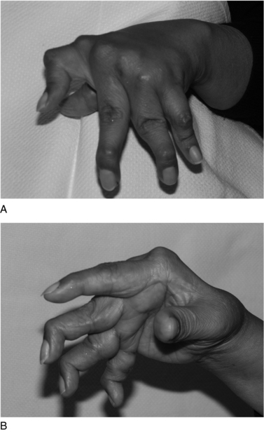
Deformities of the MP joint are usually manifested by increased ulnar drift and palmar subluxation (Fig. 17-8). The MP joint becomes unstable when normal muscle balance is lost and the collateral ligaments are disrupted from the inflammatory process. The MP joint differs from the IP joints in that its movements are not simply flexion and extension but also involve some degree of rotation, abduction, and adduction. Because of this, the MP joint is subject to greater stresses.
Ulnar drift of the MP joints is uncommon in the early stages of RA. However, Wilson44a reports 45% of patients whose disease has persisted longer than 5 years demonstrate this deformity. Ulnar drift is a combination of deviation of the phalanx from the metacarpal head and lateral shift of the phalanx upon the metacarpal.44a The following are contributors to this deformity:
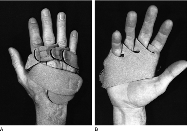
The ulnar deviation orthosis provides support to the MP joints, which restricts flexion and may assist in proper alignment by pulling the fingers radially out of ulnar deviation. The IP joints are left free to encourage proximal IP flexion during activities. This flexion may be beneficial if the patient develops swan-neck deformities. It also prevents further intrinsic tightening. This orthosis can include the wrist when it is involved. This orthosis is indicated in the acute stages of rheumatoid arthritis to decrease pain at the MP level and help prevent further deformities. It can also be used postoperatively after MP joint synovectomy to maintain flexor and extensor tendon alignment. The ulnar deviation orthosis may be used as a night splint starting 6 weeks after MP implant resection arthroplasty to assist in maintaining joint alignment (Fig. 17-9).
Thumb spica orthosis
The thumb spica orthosis may be hand or forearm based (Fig. 17-10). It provides support to the thumb CMC and MP joints. Indications may include acute rheumatoid arthritis or osteoarthritis of these joints to decrease pain and provide stability. During fabrication, the thumb should be positioned in abduction to be used as an oppositional post for the fingers. The patient should be instructed to wear the orthoses during daily activities to reduce pain and enhance function. It may be used at night to rest inflamed CMC and MP joints and provide better positioning. The orthosis may also be used postoperatively in patients who require additional stability after cast removal after trapezium or scaphoid implant resection arthroplasty. Following cast removal usually at 6 weeks, the splint is worn full-time except for bathing and gentle range of motion for 4 to 6 additional weeks.
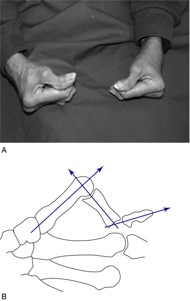
The two most common thumb deformities are the boutonnière deformity (Fig. 17-11), which is characterized by MCP joint flexion and IP joint hyperextension, and the swan neck deformity (Fig. 17-12, A), which is characterized by MCP joint hyperextension and adduction and IP joint flexion. The origin of this deformity is synovitis at the CMC joint. Severe boutonniere deformities are usually treated with an MP joint fusion in the patient with rheumatoid arthritis; however, deformities can be stabilized with a thumb spica orthosis when they are mild.
The swan-neck deformity can be treated in its mild cases or preoperatively by a long thumb spica orthosis (Fig. 17-12, B). The orthosis provides a stable post for opposition of the other fingers to the thumb, increasing function. Boutonniere deformities in which the MP joint is primarily involved can be treated with a short thumb spica splint, which does not immobilize the CMC joint and is thumb based (Fig. 17-10, B). Immobilization of the MP joint of the thumb often improves function by providing a more stable base to pinch.
Thumb interphalangeal joint orthosis
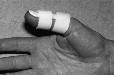
Instability and pain of the thumb IP joint [which results from OA or RA may present] in isolated deformities or in association with collapsed deformities such as swan-neck or boutonniere deformity. Immobilizing the IP joint with the use of aluminum and foam splint or small fabricated thermal plastic orthosis (Fig. 17-13) provides both stability and protection to the joint, allowing continued pinching activities without stretching of the supporting soft tissue and instability of the joint, which would make pinch difficult. The thumb IP orthosis may also be used in cases of pain and instability or when patients are considering an IP joint fusion to simulate the postoperative condition. At 6 to 8 weeks postoperatively, the splint may be used with IP joint fusions for protection following K-wire removal for an additional 2 to 6 weeks of immobilization.
Proximal interphalangeal joint orthoses
Swan-neck and boutonniere deformities are the most common finger deformities.33a Synovitis of the PIP joint produces a painful, swollen joint. If synovitis continues it spreads proximally under the central extensor slip, which becomes attenuated, and between the extensor and intrinsic tendons. When the central slip and lateral bands stretch, they migrate volarly and a boutonniere deformity is produced.33a,44a Nalebuff has described three stages of the boutonniere deformity.28a Stage I exhibits only a slight extensor lag, and a slight loss of DIP flexion occurs. A 40 degree PIP flexion deformity is considered stage II. In stage III, the PIP joint has a fixed flexion deformity.
If the initiating PIP synovitis is anterolateral, it can stretch the transverse retinacular ligament, allowing the lateral band to migrate dorsally, resulting in a PIP joint hyperextension, or swan-neck deformity. Lateral bands in this position prevent the normal volar and lateral shift that allows PIP flexion. With progression of the synovitis, joint erosion and finally destruction occur with fibrous articular adhesions or bony ankylosis.45 The swan-neck deformity can also be caused by the destructive effect of the synovitis beginning at any one of the three digital joints (Rizio & Belsky).46
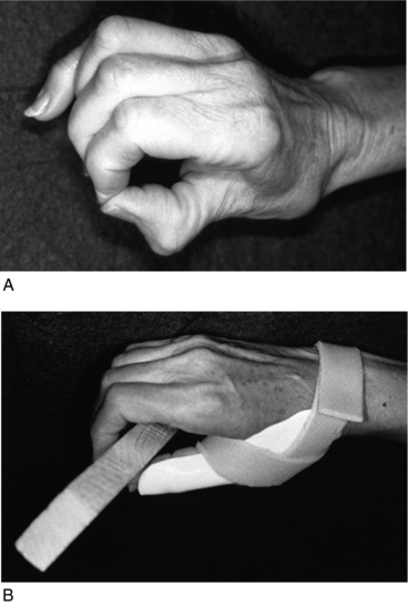
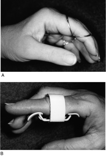
Preoperatively a static orthosis may be fabricated to prevent swan-neck or boutonniere deformities from becoming fixed and to improve hand function. A commonly used orthosis for this situation is a figure-of-eight ring orthosis (Fig. 17-14, A) or tripoint finger splint (Fig. 17-14, B, and Fig. 17-5, B). The orthosis applies pressure at three points on the finger, providing a correcting force opposite to that of the deformity. For example, in splinting the swan-neck deformity, the pressure is applied dorsally, proximally, and distally, to the PIP joint and volarly over the center of the joint. This results in a flexion force at the PIP joint, which consequently presents with PIP hyperextension and allows active PIP flexion. This splint may be used preoperatively and postoperatively. However, it is not recommended in the immediate postoperative period because of swelling. A dorsal PIP splint is better immediately postoperatively because it is less constricting. Prevention of hyperextension by splinting in flexion allows structures volar to the PIP joint to shorten and help prevent return of the deformity. For the boutonniere deformity, the orthosis is applied opposite that of the swan-neck deformity. There is pressure over the central aspect of the PIP joint dorsally and proximal and distal to the PIP joint volarly resulting in an extension force preventing PIP flexion contractures.
< div class='tao-gold-member'>
Stay updated, free articles. Join our Telegram channel

Full access? Get Clinical Tree


