Opening Wedge Tibial Osteotomy


Opening Wedge Tibial Osteotomy


Frank Noyes


CONTRAINDICATIONS

HTO is contraindicated in knees in which there is more than a 15 by 15 mm area of exposed bone on both the tibial and the femoral surfaces. There are younger patients in whom the area of exposed bone may be larger and partial knee replacement is not an option. However, as a general rule, articular cartilage should be present over the majority of the medial joint surfaces. The difficult decision is the treatment recommendation for patients between 50 and 60 years of age. With the increasing longevity of unicompartmental knee replacements, patients who have advanced medial compartment damage with major areas of bone exposure will likely experience symptoms after HTO and are better candidates for partial knee replacement.

Major concavity of the medial tibial plateau with loss of bone stock is a contraindication to HTO. Knees that demonstrate (on standing 45° posteroanterior radiographs) no remaining articular cartilage space in the medial compartment are not candidates. Additional contraindications are a limitation of knee flexion (>10°), lateral tibial subluxation (>10 mm), prior lateral meniscectomy, or lateral tibiofemoral joint damage.

An absolute contraindication for a medial opening wedge osteotomy is the use of nicotine products in any form. A relative contraindication is obesity (body mass index >30) because unloading of the medial compartment will not be achieved. Another relative contraindication is increased medial slope to the affected medial tibial plateau
in the coronal plane due to advanced medial plateau concavity. This finding indicates that it will not be possible to significantly unload the medial compartment, and a majority of the weight-bearing loads will be confined to the medial compartment. Marked patellofemoral symptoms contraindicate an HTO. Medical contraindications include diabetes, rheumatoid arthritis, autoimmune diseases, and malnutrition states.


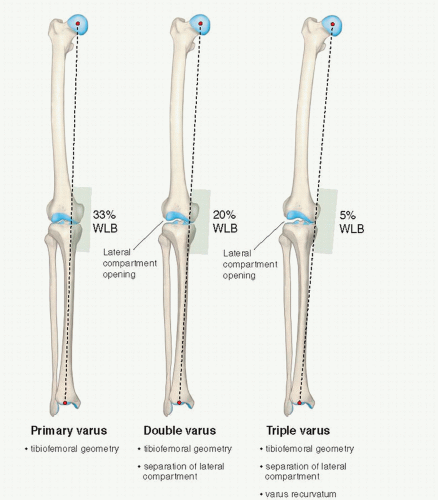
FIGURE 76.1. Schematic illustration of primary, double, and triple varus knee angulation. WBL, weight-bearing line. (Reprinted from Noyes FR, Barber-Westin SD. Primary, double, and triple varus knee syndromes: diagnosis, osteotomy techniques, and clinical outcomes. In: Noyes FR, ed. Noyes Knee Disorders: Surgery, Rehabilitation, Clinical Outcomes. Philadelphia, PA: Saunders; 2009:821-895.)


CLINICAL EVALUATION

Patients complete questionnaires and are interviewed for the assessment of symptoms, functional limitations, sports and occupational activity levels, and patient perception of the overall knee condition according to the Cincinnati Knee Rating System (5) or other validated knee rating instruments.

The physical examination of the knee joint to detect all of the abnormalities in the varus-angulated knee includes assessment of (1) the patellofemoral joint, especially extensor mechanism malalignment due to increased external tibial rotation and posterolateral tibial subluxation; (2) medial tibiofemoral crepitus on varus loading, indicative of articular cartilage damage; (3) pain and inflammation of the lateral soft tissues due to tensile overloading; (4) gait abnormalities (excessive hyperextension or varus thrust) during walking and jogging (6); and (5) abnormal knee motion limits and subluxations compared with the contralateral knee (7).


The medial posterior tibiofemoral step-off on the posterior drawer test is done at 90° of flexion. This test is performed first to determine that the tibia is not posteriorly subluxated, indicating a partial or complete posterior cruciate ligament (PCL) tear. The Lachman and pivot-shift tests are performed. FCL insufficiency is determined by the varus stress test at 0° and 30° of knee flexion. An increase in medial joint opening may occur compared with the opposite knee that represents a pseudolaxity, as the increase is actually due to medial tibiofemoral joint narrowing. The true amount of medial and lateral tibiofemoral compartment opening is later confirmed during the arthroscopic examination with gap tests (Fig. 76.2).


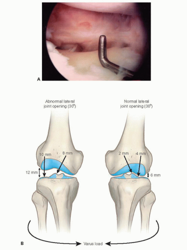
FIGURE 76.2. Arthroscopic gap test for determining the amount of lateral joint opening. (Reprinted from Noyes FR, Barber-Westin SD. Primary, double, and triple varus knee syndromes: diagnosis, osteotomy techniques, and clinical outcomes. In: Noyes FR, ed. Noyes Knee Disorders: Surgery, Rehabilitation, Clinical Outcomes. Philadelphia, PA: Saunders; 2009:821-895.)

The tibiofemoral rotation dial test (8) is used to estimate the amount of posterior tibial subluxation. A varus recurvatum test in both the supine and the standing positions as well as the reverse pivot shift test are included in the assessment of posterolateral tibial subluxation.

Radiographic assessment of lower limb alignment is based on double-stance, full-length anteroposterior radiographs showing both lower extremities (knee flexed 3° to 5°) from the femoral heads to the ankle joints (9).
If separation of the lateral tibiofemoral joint is observed, it is necessary to subtract the lateral compartment opening so that the true tibiofemoral osseous alignment is determined and a valgus overcorrection is avoided. Other radiographs include a lateral at 30° knee flexion, weight-bearing posteroanterior at 45° knee flexion, and patellofemoral axial views. Telos medial or lateral stress radiographs may also be required of both knees. The height of the right and left patella is measured on lateral radiographs to determine if an abnormal patella infera or alta position exists (1).


PREOPERATIVE PLANNING

The preoperative calculations for HTO involve precise measurements to determine the amount of angular correction desired to redistribute tibiofemoral forces, whereas not altering tibial slope and tibiofemoral joint obliquity in the frontal plane (Table 76.1) (1, 9, 10).


Table 76.1 Preoperative planning


Determine angular correction to achieve redistribution of tibiofemoral forces without altering tibial slope. Take into account abnormal lateral tibiofemoral joint separation from deficient posterolateral structures. Measure WBL on bilateral standing hip-knee-ankle radiographs:


WBL is dependent on femoral and tibial lengths and angular deformity


Measure tibial slope on lateral radiographs.


Increasing tibial slope increases anterior tibial translation; potentially, tensile loads on the ACL.


Decreasing tibial slope increases posterior tibial translation; potentially, tensile loads on PCL.


Do not alter normal tibial slope unless it is markedly abnormal:


Tibial slope greater than two standard deviations above normal


Do not alter a normal tibal slope in ACL-deficient or PCL-deficient knees.


Maintain normal tibial slope: anterior gap at medial opening wedge should be one-half the posteromedial gap.


Every 1 mm of anterior gap change = 2° change in tibial slope.


Calculation of millimeters opening posteromedial tibial cortex based on law of triangles for coronal alignment


correction.


Timing of HTO in knees with ligament deficiencies:


Primary varus knees:


Cruciate reconstruction with HTO or later (no abnormal lateral joint opening present)


Double varus knees:


HTO first


Posterolateral structures may shorten with valgus alignment


Perform cruciate, posterolateral reconstructions later if required


Triple varus knees:


HTO first, cruciate and posterolateral reconstruction later


Opening wedge osteotomy advantages:


Avoids lateral dissection, fibular osteotomy


Large correction >12°, avoids tibial shortening


Distal advancement or reconstruction of the MCL in chronic MCL ruptures


In subsequent posterolateral reconstructions, avoids proximal fibular osteotomy, allows FCL grafts to be fixated securely to proximal fibula


From Noyes FR, Barber-Westin SD. Primary, double, and triple varus knee syndromes: diagnosis, osteotomy techniques, and clinical outcomes. In: Noyes FR, ed. Noyes Knee Disorders: Surgery, Rehabilitation, Clinical Outcomes. Philadelphia, PA: Saunders; 2009:821-895.


An under- or overcorrection in the coronal plane may result if the surgeon fails to recognize the effect of lateral tibiofemoral joint separation on increasing varus angulation that results from slack or deficient lateral soft tissues. Two methods are used to determine the correction wedge on preoperative radiographs, which have been described in detail elsewhere (Figs. 76.3 and 76.4) (9). Lateral radiographs are examined and measurements made of the tibial slope (10, 11).

There are patients who have a distinctly abnormal tibial slope from a prior osteotomy or tibial fracture, or growth abnormality where correction of the tibial
slope is required before cruciate ligament surgery or other conditions discussed. Empirically, a tibial slope greater than two standard deviations above normal (e.g., a tibial slope of 15° or greater) usually requires correction.


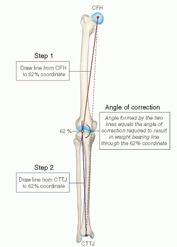
FIGURE 76.3. Graphic depiction of the method used to calculate the correction angle of an HTO using a full-length anteroposterior radiograph of the lower extremity. The lines from the centers of the femoral head (CFH) and tibiotalar joint (CTTJ) converge in this example at the 62% coordinate. (Reprinted from Noyes FR, Barber-Westin SD. Primary, double, and triple varus knee syndromes: diagnosis, osteotomy techniques, and clinical outcomes. In: Noyes FR, ed. Noyes Knee Disorders: Surgery, Rehabilitation, Clinical Outcomes. Philadelphia, PA: Saunders; 2009:821-895.)

The rule to remember is that the anterior gap at the medial opening wedge should be one-half of the posteromedial gap to maintain a normal tibial slope (11). For every 1 mm of anterior gap change, an approximate 2° change in tibial slope would be produced (Fig. 76.5). This is based on the angle of the anteromedial tibial cortex, tibial width, and the Anteroposterior (AP) distance where the gap measurement is made. The millimeters of opening of the posteromedial tibial cortex is based on the law of triangles (Table 76.2) and confirmed at surgery. The surgeon should determine the proper gap width of the osteotomy opening wedge along the anteromedial cortex to maintain tibial slope and the proper width beneath the tibial plate based on its location along the anteromedial cortex (Table 76.3). The opening wedge gap will always be 3 to 4 mm less where the plate is located.

The timing of HTO and ligament reconstructive procedures is based on several factors discussed elsewhere (Fig. 76.6) (1). The author prefers to perform the HTO first and, after adequate healing of the osteotomy, the required ligament reconstructive procedures. The preferred grafts and operative techniques for ACL (12), PCL (13) and posterolateral ligament (14) reconstructions are described elsewhere.


OPERATIVE TECHNIQUE: OPENING WEDGE TIBIAL OSTEOTOMY

All knee ligament subluxation tests are performed after the induction of anesthesia in both the injured and the contralateral limbs. A thorough arthroscopic examination is conducted, documenting articular cartilage surface abnormalities and the condition of the menisci. The gap test is done during the arthroscopic examination. Knees that have 12 mm or more of joint opening at the periphery of the lateral tibiofemoral compartment will usually require a staged posterolateral reconstructive procedure. Associated meniscus tears are either repaired if possible (15) or partially removal. Appropriate debridement of tissues, inflamed synovium, and notch osteophytes limiting knee extension is performed.

Preoperative calculations are made as previously described. The entire lower extremity is prepped and draped free with the tourniquet placed high on the proximal thigh to assist visual observation of lower limb alignment. If an autogenous iliac crest autograft is to be performed (authors’ choice), the ipsilateral anterior iliac crest is prepped and draped for the limited iliac crest bone harvest of the outer cortex.

The technique for the opening wedge osteotomy is summarized in Table 76.4 and has been described in detail elsewhere (1). The iliac crest bone harvest involves a 4-cm incision made over the anterior iliac crest and deepened to the periosteum (Fig. 76.7A and B). In most patients, the graft will be 40 mm in length, 10 mm in width, and 30 mm in depth. However, in smaller patients, the graft may be smaller in width, approximately 8 mm in depth. Patients undergoing large osteotomies may require a longer graft of approximately 45 to 50 mm. The inner iliac cortex is not dissected, the muscle attachments are not disturbed, which reduces postoperative pain, and a spacer of the iliac crest defect is not required.

The operative technique is shown in Figure 76.8. A 5-cm vertical skin incision is made medially midway
between the tibial tubercle and the posteromedial tibial cortex, starting 1-cm inferior to the joint line. Once the dissection is complete, a Keith needle is placed in the anteromedial joint just above the tibia, and the distance is marked on the desired point of the osteotomy along the anteromedial cortex. A second Keith needle is placed at the posteromedial tibial joint space, and the same millimeters are marked to provide a measurement of the tibial slope. The two marks are connected to provide the osteotomy line perpendicular to the tibial slope.


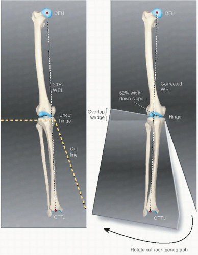
FIGURE 76.4. Graphic depiction of an alternative method used to calculate the correction angle of an HTO using a full-length anteroposterior radiograph of the lower extremity. The roentgenograph is cut to allow the center of the femoral head (CFH), the 62% coordinate, and the center of the tibiotalar joint (CTTJ) to become colinear. The angle of the resulting wedge of roentgenograph overlap equals the desired angle of correction. The example is provided for a closing wedge osteotomy. The same technique is used for an opening wedge osteotomy where the medial tibial opening wedge is made to obtain the desired correction. (Reprinted from Noyes FR, Barber-Westin SD. Primary, double, and triple varus knee syndromes: diagnosis, osteotomy techniques, and clinical outcomes. In: Noyes FR, ed. Noyes Knee Disorders: Surgery, Rehabilitation, Clinical Outcomes. Philadelphia, PA: Saunders; 2009:821-895.)

Only gold members can continue reading. Log In or Register to continue

Stay updated, free articles. Join our Telegram channel

Jul 22, 2016 | Posted by in ORTHOPEDIC | Comments Off on Opening Wedge Tibial Osteotomy

Full access? Get Clinical Tree

Get Clinical Tree app for offline access