Open Reduction Internal Fixation of the Ulnar Styloid
Evan D. Schumer
Bruce M. Leslie
Indications and Contraindications
The association between ulnar styloid fractures and distal radius fractures has been noted for many years, but its significance has not been conclusively shown. Frykman’s eponymous fracture classification implied that the presence of a distal ulnar styloid fracture meant a higher, more complex classification level, but did not provide guidance in terms of treatment. Recent depictions of the ligamentous attachments about the ulnar head have shown that most of the ligaments originate from the fovea and the base of the ulnar styloid; the styloid is usually devoid of major ligaments (1).
The oft-quoted Knirk and Jupiter (2) article noted that 31 of 43 intra-articular fractures of the distal radius had an associated ulnar styloid fracture. Of the 31 ulnar styloids, 20 developed nonunion; 9 of the 11 that healed had good to excellent results, whereas only 10 of the 20 nonunions had good to excellent results (2).
Why is this seemingly trivial fracture of the ulnar styloid, the distal aspect of which has no ligamentous attachments., historically led to such poor clinical results (3)? Perhaps the presence of the fracture itself signifies a more complex ligamentous injury on the ulnar side of the wrist. Perhaps its mere presence should alert the clinician to the possibility of distal radioulnar joint (DRUJ) instability, and the need to correct this problem if it exists at the time of repair of the distal radius fracture.
Open reduction internal fixation should not be performed in the face of grossly contaminated wounds. It should be used judiciously in the face of clean open fractures as long as there is adequate soft-tissue coverage. Osteoporotic bone is a relative contraindication. Techniques that do not depend on screw fixation may be more effective with osteoporotic bone.
Open reduction internal fixation requires the use of intraoperative radiography and consequently should not be undertaken without ready access to a fluoroscopy unit.
Preoperative Preparation
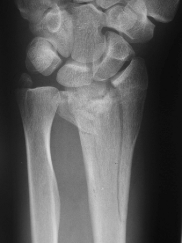
No clear definitive indications exist as to which ulnar styloid fractures should be repaired, but it is generally accepted that ulnar styloid fractures associated with DRUJ instability should be reduced and stabilized. Instability should be suspected if preoperative radiographs demonstrate a displaced sigmoid notch fracture involving the dorsal or palmar ridge (Fig. 29-1), or a markedly displaced ulnar styloid fracture (Fig. 29-2) (4).
Moore et al. (5) offered four radiographic signs of DRUJ capsule and triangular fibrocartilage complex (TFCC) rupture:
Ulnar styloid base fracture with more than 2 mm of displacement
Widening of the DRUJ base on anteroposterior (AP) view

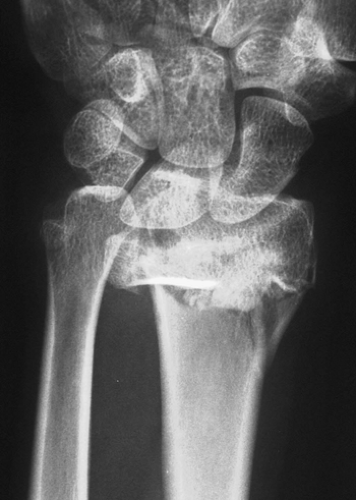
Figure 29-2 Comminuted intra-articular distal radius fracture with displaced base of ulnar styloid fragment.
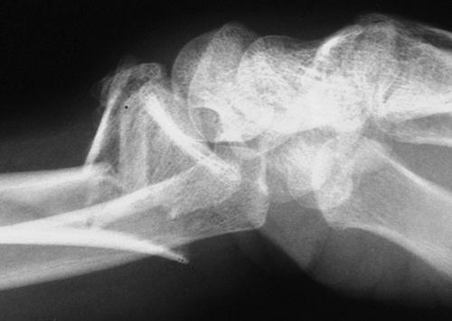
Dislocation on the lateral view (Note: This can be difficult to assess owing to the shape of the distal ulna and computerized tomography [CT] scan of both forearms in equal amounts of rotation for comparison is the test of choice to confirm) (Fig. 29-3).
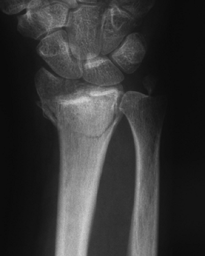
More than 5 mm of radial shortening (Fig. 29-4).
This suspicion for instability should prompt an examination of the unaffected wrist in neutral, pronation, and supination to judge the ligamentous integrity of the intact wrist preoperatively (5).
Evaluate stability of the DRUJ in the injured wrist by manipulating the distal ulna in a dorsal-volar direction in all positions of rotation (Fig. 29-5A, B) after stabilizing the distal radius fracture. Compare the fractured wrist with the uninvolved wrist. With obvious laxity, stabilize the DRUJ. The chosen technique will depend on the radiographic appearance, patient demographics, and equipment available, as well as the experience of the surgeon.
In recent years, newer more anatomic and rigid fixation systems have been introduced to address distal radius fractures. Frequently with these techniques, sigmoid notch fragments are successfully reduced and rigidly held, and radial height is restored. Both of these factors are likely to result in increased stability of the DRUJ after radius fixation alone. In some instances, the ulna styloid fragment is seen to fall back toward its anatomic position with these newer techniques (Figs. 29-6 and 29-7).
Technique
In pronation, the ulnar styloid will be on the ulnar side of the wrist just volar to the extensor carpi ulnaris tendon (Fig. 29-8). The dorsal sensory branch of the ulnar nerve lies just distal to the ulnar styloid as it passes onto the dorsum of the hand, and is likely to be in the operative field for any approach to the ulnar styloid.

Figure 29-3 Apparent volar dislocation of ulnar head relative to dorsally displaced distal radial fragments on lateral radiograph.

Figure 29-4 Comminuted distal radius fracture demonstrating more than 5 mm of radial shortening compared with the intact ulna.
Perhaps the most straightforward approach to the ulna styloid is to expose the DRUJ beneath the extensor carpi ulnaris (ECU) subsheath. Create a dorsal ulnar skin incision; blunt dissection allows the dorsal sensory branch of the ulnar nerve to be identified and protected.
Stay updated, free articles. Join our Telegram channel

Full access? Get Clinical Tree


