Open Reduction and Internal Fixation with a Volar Locking Plate
Mohana Amirtharajah
Andrew Weiland
Indications
Fractures of the distal radius are among the most common conditions encountered by the practicing orthopaedic surgeon. Although minimally displaced fractures can still be addressed by closed means, anatomic restoration of the distal radius is the goal of the treating physician. Open reduction and internal fixation with volar locked plating is an essential tool in the treatment of distal radius fractures. Volar locked plating provides secure restoration of articular congruency, allowing early motion with minimal soft tissue and tendon trauma.
Volar locked plating can be used to achieve near-anatomic reduction of the distal radius. Generally accepted guidelines for reduction include radial shortening less than 5 mm, radial inclination greater than 15 degrees, sagittal tilt between 5 degrees of dorsal and 20 degrees of volar tilt, and articular congruity with less than a 2-mm step-off at the radiocarpal and radioulnar articulations (1). Although these goals can be achieved by pins and plaster and/or external fixation, these methods rely on indirect ligamentotaxis to control the fracture fragments. In addition, external fixation can be complicated by extrinsic tightness of the digital extensors, wrist stiffness, malunion, and complex regional pain syndrome (2). Dorsal buttress plating is also a useful technique since many fractures of the distal radius tend to collapse dorsally. However, even with newer low-profile designs, extensor tendon tenosynovitis, attrition, and rupture remain concerns (3,4). Furthermore, biomechanical studies have suggested that volar fixed-angle plating has increased rigidity and load to failure with axial loads compared to other plating techniques (5).
For these reasons, volar fixed-angle plating has become the workhorse in the treatment of distal radius fractures. This technique uses a fixed-angle plate and screw construct to support the subchondral bone and articular fragments. Indications include displaced fractures with a large potential to collapse or with articular comminution that cannot be acceptably corrected by closed means. Both apex volar and apex dorsal as well as intra- and extra-articular fractures can be treated in this manner. This technique is particularly useful in osteopenic or metaphyseally deficient bone (2). Finally, volar fixed-angle plating is also indicated when early motion is desired.
Contraindications
Although the optimal use of volar plating is still being established, there are few true contraindications. They include skeletally immature patients with open distal radial physes. Its use in patients with inadequate volar soft-tissue coverage for the plate or those in whom an active infection is proven or suspected is also contraindicated. Often, extremely distal fractures with a very thin subchondral fragment and accompanying dislocation of the carpus are not treated adequately by volar plating
alone (2). Dorsal shear-type fractures (dorsal Barton’s) also are not treated ideally by this means. In addition, there have been reports of failure in patients with a volar shear fracture of the lunate facet after adequate initial reduction, suggesting that supplemental fixation may be necessary in this case (6). Finally, fractures that extend into the radial diaphysis can be treated only with volar locked plating if a long enough plate is available (2).
alone (2). Dorsal shear-type fractures (dorsal Barton’s) also are not treated ideally by this means. In addition, there have been reports of failure in patients with a volar shear fracture of the lunate facet after adequate initial reduction, suggesting that supplemental fixation may be necessary in this case (6). Finally, fractures that extend into the radial diaphysis can be treated only with volar locked plating if a long enough plate is available (2).
Preoperative Preparation
A detailed history is ascertained in order to document the mechanism of injury and the amount of energy imparted into the fracture site. In addition, a careful neurologic exam should be undertaken to rule out possible nerve compression or compartment syndrome. The integrity of the tendons, particularly the extensor pollicis longus, should be well documented. Moreover, the condition and integrity of the soft tissues should be carefully assessed. Although patients with a great deal of swelling and ecchymosis can tolerate a volar incision, the presence of blistering or skin necrosis suggests a significant soft-tissue injury that may need an extended period of strict elevation and reassessment of the soft tissues prior to proceeding. Finally, elderly patients are interviewed carefully to determine whether their fall was secondary to an underlying medical condition that may warrant further work-up or consultation with a medical, cardiac, or neurologic service.
Initial radiographic evaluation includes routine posteroanterior (PA), lateral, and oblique radiographs of the wrist. Initial injury films are critical in determining the amount and direction of initial displacement (Fig. 5-1). Traction and/or postreduction films are also important in defining the fracture fragments. Radiographs of the forearm, elbow, hand, or scaphoid are taken if clinically indicated. Careful evaluation of carpal alignment is also necessary. Although routine computed tomography for distal radius fractures is not performed, advanced imaging may be necessary to further delineate the fracture fragments, evaluate the integrity of the lunate facet, determine the extent of comminution and the thickness of the subchondral fragment, and ascertain the presence of a dorsal shear fragment.
Technique
Surgery is almost always performed under regional anesthesia. All patients receive antibiotic prophylaxis prior to surgery.
Apply a nonsterile tourniquet to the upper arm.
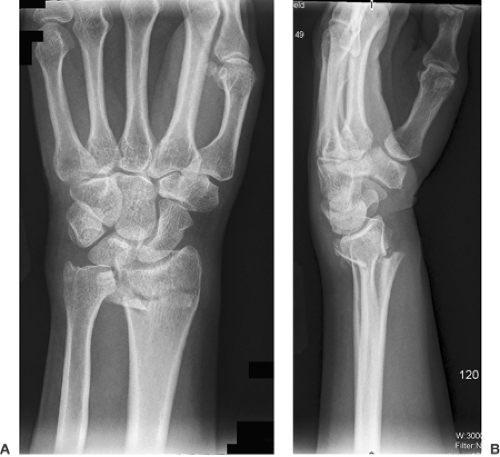
Figure 5-1 Posteroanterior and lateral views of the wrist show a shortened and dorsally angulated fracture of the distal radius.
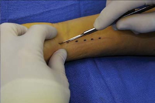
Figure 5-2 Make an incision over the flexor carpi radialis tendon.
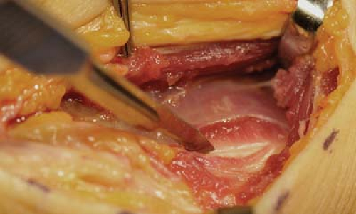
Figure 5-3 Visualize the pronator overlying the distal radius and incise it with a cuff of muscle left for later repair.
Exsanguinate the limb and inflate the tourniquet to 250 mm Hg.
Make a 5-cm incision over the flexor carpi radialis (FCR) tendon (Fig. 5-2). If more exposure is needed, turn the distal limb radially at a 45-degree angle to the wrist crease.
Continue the incision down through skin and subcutaneous tissue, paying careful attention to protecting the palmar cutaneous branch of the median nerve.
Identify the FCR tendon sheath, and incise it on its radial border.
Retract the FCR tendon ulnarly, and incise the floor of the tendon sheath.
Retract the flexor pollicis longus tendon ulnarly along with the flexor digitorum superficialis tendons, the flexor digitorum profundus tendons, and the median nerve. The pronator quadratus is visible as it lies over the distal radius. Often with high-energy fractures, this muscle is lacerated. However, its remnants may be subperiosteally elevated from the underlying radius by making an L-shaped incision over its insertion distally and radially (Fig. 5-3).
When removing this muscle from its distal extent, be careful not to enter the radiocarpal joint or damage the palmar carpal branch of the radial artery, which can be found in this region.
Leave a small sleeve of tissue is radially and distally for later repair.
Lift the pronator off the radius until there is enough exposure proximally and distally for screw placement (Fig. 5-4).Related posts:

Stay updated, free articles. Join our Telegram channel

Full access? Get Clinical Tree


