Open Fractures
Mark A. Hardy
Jordan P. Grossman
An open fracture (Fig. 96.1) is characterized by soft tissue disruption that results in communication of the fracture site with the outside environment (1,2 and 3). Open fracture management includes meticulous wound débridement, tetanus prophylaxis, early and appropriate antibiotic therapy, and fracture stabilization. The treatment objectives, simply stated, are prevention of wound sepsis, obtaining fracture healing, and restoring function.
EVALUATION, PATHOMECHANICS, AND RATIONALE
Once Advanced Trauma Life Support protocols have been met, the injured extremity is often one of the patient’s most significant injuries (1). Evaluation and treatment of these injuries include not only the fracture itself but also the precarious soft tissue structures such as the neurovascular structures, tendons, and ligaments.
GUSTILO-ANDERSON CLASSIFICATION
To help standardize care and comparison of similar injuries in studies, Gustilo and Anderson (2) classified open fractures into three categories (Table 96.1; Fig. 96.2). Gustilo further stratified type III wounds according to a worse prognosis with a wound sepsis rate (Table 96.2).
The Gustilo-Anderson classification is somewhat prognostic in that its treatment protocols parallel the antibiotics recommended. However, since the system is based on tibial shaft fractures, it does have some limitations in its translation to injuries of the foot and ankle. Additionally, in a study by Brumback and Jones (4), there was found to be only a 60% observer agreement in classification of tibial fractures. It was found that the majority of these misclassifications occurred with relatively small skin wounds, such as seen with gunshot wounds (Fig. 96.3). Therefore, to improve classification accuracy, the extent and severity of the injury should be assessed during surgery, after thorough wound exploration and débridement.
RADIOGRAPHS
Plain-film radiographs taken in various cardinal planes are obtained as appropriate to the anatomic site of injury (Fig. 96.4). Advanced imaging, such as computed tomography radiography, is often required when there is an intra-articular component or in cases in which there is significant comminution.
WOUND CULTURES
Controversy exists in the usefulness of wound cultures in open fractures and dislocations. This is due to the failure of cultures either taken in the emergency room or predébridement to identify the infection causing organism. In a randomized study by Patzakis et al (5), they demonstrated that only 18% of all infections that occurred in a series of 171 open fractures were caused by an organism identified by the initial wound culture. This points to the fact that most infections in open injuries are the result of nosocomial organisms and stresses the importance of early wound coverage.
TREAT AS EMERGENT/GOLDEN PERIOD
Open fractures have long been considered emergencies. The American College of Surgeons Committee on Trauma, in its Resources for Optimal Care of the Injured Patient, indicates 6 hours as the benchmark for time from injury to débridement of open fractures in trauma centers (6). This is based on studies that have shown that inoculation of 1 g of tissue with a single bacterium will replicate 105 bacteria within 6 hours, and this is clinically defined as an infection.
COMPARTMENT SYNDROME
Although rare, compartment syndrome should not be overlooked in the management of open injuries of the foot and ankle. The open fracture compartment may indeed be spared but adjacent compartments remain at risk. The surgeon should maintain a low threshold for fasciotomy and an even lower threshold for measurement of the compartment pressures. Generally speaking, any compartment with pressures greater than 30 mm Hg requires decompression (Fig. 96.5).
INDICATIONS FOR AMPUTATION
Occasionally, the decision must be made whether to attempt limb salvage or perform a primary limb amputation (Fig. 96.6). There are multiple factors that must be considered in this decision. To aid the surgeon, several scoring systems have been developed. Each system indentifies host variables and assigns them a value. These variables may include such things as limb ischemia, nerve injury, patient age, shock, and the degree of skeletal and soft tissue injury. Some of the more commonly used scoring systems are the Mangled Extremity Severity Score, Predictive Salvage Index, Limb Salvage Index, and Hannover Fracture Scale-97.
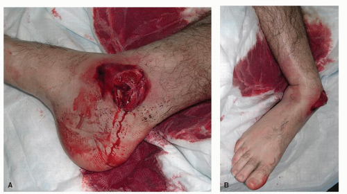 Figure 96.1 Grade III open distal tibial fracture (A) with significant dislocation (B). |
SURGICAL TECHNIQUE
DÉBRIDEMENT
Thorough surgical débridement plays a critical role in the management of open fractures because devitalized tissue and foreign material promote the growth of microorganisms and constitute a barrier for the host’s defense mechanisms (3). When possible, a tourniquet should be avoided so the surgeon can better evaluate the integrity of the bone and soft tissues.
IRRIGATION
Along with débridement, irrigation is believed to be the most important factor in reducing the prevalence of infection. Despite this, there is little scientific research concerning the factors that influence irrigation (7,8). Normal saline is the solution most often used. Although, some have advocated the addition of antibiotics or antiseptics to the solution, no definitive studies have shown this to be beneficial. It is the mechanical properties of the irrigant that reduce the incidence of infection rather than its chemical properties (Fig. 96.7). Traditionally, the irrigation fluid has been delivered via a bulb syringe and aided by a scrub brush and suction; however, there are many pulse
lavage systems that are now available. Advocates of the pulsed lavage system claim that it works more effectively to remove contaminants from wounds than the bulb syringe (9,10). In a study by Bhandari et al (11), they showed that microscopic damage to the bony architecture could be caused by pulsed lavage; therefore, a pressure threshold of 50 psi was identified, beyond which new bone formation was inhibited.
lavage systems that are now available. Advocates of the pulsed lavage system claim that it works more effectively to remove contaminants from wounds than the bulb syringe (9,10). In a study by Bhandari et al (11), they showed that microscopic damage to the bony architecture could be caused by pulsed lavage; therefore, a pressure threshold of 50 psi was identified, beyond which new bone formation was inhibited.
TABLE 96.1 Gustilo-Anderson Classification of Open Fractures | ||||||
|---|---|---|---|---|---|---|
|
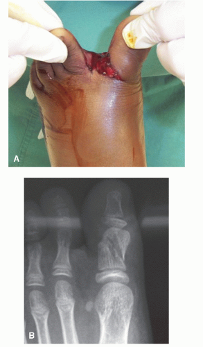 Figure 96.2 A: Grade I open pediatric hallux fracture. B: Note the simple fracture pattern with viable soft tissue envelope. |
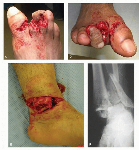 Figure 96.2 (Continued) C,D: Grade II open forefoot injury secondary to a lawnmower accident. Note complete and near complete amputations of the second and third digits, respectively. E,F: Grade III open trimalleolar ankle fracture sustained after a fall from a height. |
TABLE 96.2 Gustilo’s Classification of Type III Open Wounds | ||||||
|---|---|---|---|---|---|---|
|
ANTIBIOTIC THERAPY
The efficacy of antibiotics in open fractures is well accepted—to the point that they are no longer considered prophylactic but rather therapeutic. Antibiotic therapy should adhere to the following principles (12,13):
Related posts:

Stay updated, free articles. Join our Telegram channel

Full access? Get Clinical Tree


