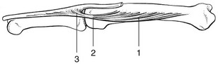
of the hand and fingers


Disorders of the hand and fingers


image


Pain and paraesthesia are two symptoms that are common in the hand. Pain is very often the result of either local trauma or overuse. Precise localization is possible in pain that is not referred from a lesion higher up in the limb. Paraesthesia may reflect a proximal lesion and the patient has difficulty in identifying the source of his symptoms.


Disorders of the inert structures


The capsular pattern


Any of the joints of the fingers may become affected by one or other form of arthritis, which results in limitation of movement with a capsular pattern.


The capsular pattern at a finger joint is an equal loss of movement at the beginning and end of the normal range in either direction. Some movement remains possible at the midpoint. Rotations are painful at the extremes of range. In very severe arthritis, rotations may also become limited.


The presence of a capsular pattern indicates that an arthritis has developed, the type of which can be defined from the history. The following questions are relevant:


Rheumatoid arthritis


Rheumatoid arthritis is undoubtedly the most deforming and most incapacitating disorder of the hand.1


The symptoms and signs usually develop in the course of a few weeks or months and are often symmetrical. Frequently, the first symptom is morning stiffness of the fingers. The basic functional examination is usually negative, but tenderness to touch can be elicited. The erythrocyte sedimentation rate is markedly raised.


Early in the course, a capsular pattern develops and one or more metacarpophalangeal joints or proximal interphalangeal joints of one or both hands show the familiar spindle-shaped swelling.


Later, when osseous destruction takes place, a palmar luxation of the fingers occurs and they deviate towards the ulnar side as the result of subluxation in the metacarpophalangeal joints. The fingers may develop the typical buttonhole and swan neck deformities: the former results in hyperextension of the metacarpophalangeal joint, flexion at the proximal interphalangeal joint and extension at the distal interphalangeal joint; the latter results in flexion at the metacarpophalangeal joint, hyperextension at the proximal and flexion at the distal interphalangeal joints. The thumb becomes Z-shaped (typical ‘ninety-ninety’ deformity): the metacarpophalangeal joint is fixed in 90° flexion, the interphalangeal joint in 90° extension. The joints are also warm to the touch.


Secondary rupture of tendons and ligaments may occur, with subsequent muscular contractures or muscular atrophy.


Treatment is systemic. Only in the initial stage, when few joints are affected, may intra-articular triamcinolone prove effective.


As in any other patient, symptoms may develop that have nothing to do with the patient’s rheumatoid arthritis. Trigger finger, carpal tunnel syndrome and de Quervain’s disease are common, and another possibility is a ganglion lying between the heads of the second and third metacarpal bones, which gives rise to vague local aching and responds well to aspiration.2


Traumatic arthritis


The typical history of direct contusion, indirect sprain or reduced dislocation of a finger joint indicates the presence of a traumatic arthritis – a very common condition. A chip fracture may also be responsible for the arthritis.


On inspection, a spindle-shaped swelling is often seen, which resembles the swelling of rheumatoid arthritis. Examination further reveals a capsular pattern, and on palpation warmth may be felt, especially after a severe injury. As the arthritis may be combined with a tendinous lesion, resisted movements of the fingers must also be tested.


Traumatic arthritis of the finger joints does not respond satisfactorily to any treatment. Intra-articular injection with a steroid, so useful in traumatic arthritis in the toe joints, affords no corresponding benefit in the fingers. Recovery is spontaneous over 6–18 months, depending on the severity of the original trauma and the age of the patient. Sometimes manual therapeutic techniques may favourably alter the natural course.3 Immobilization is strongly contraindicated.


Arthrosis


Occasionally arthrosis in one joint develops as the result of severe injury but more often the condition has a spontaneous onset and affects several joints. Women between 40 and 60 years of age are often affected, and there is a strong familial predisposition.4


Arthrosis begins at the distal interphalangeal joints, and its knobbly appearance is quite different from rheumatoid arthritis. Both hands are usually affected more or less symmetrically. The index, middle and ring fingers are most usually affected. At the base of the distal phalanx, two small rounded bosses on the dorsum of the joint (Heberden’s nodes)5 can be seen. A varus deformity may develop at a distal joint, usually at the index. Some years later, the arthrosis may spread to the proximal interphalangeal joints (with the formation of nodes at index and middle fingers – Bouchard’s nodes); it seldom reaches the metacarpophalangeal joints. From time to time, a new node forms at an affected joint and the patient will mention some aching or slight pain over 1 or 2 months, during which time the fingertip may occasionally become pink. The colour is mottled and different from the shiny red of gout. After a month or two the discolouration passes off and the node ceases to be painful.


As the joints of the hand are not weight-bearing, arthrosis is not a very painful condition. The patient merely complains of stiffness and aching especially after exertion.


The radiograph clearly shows the usual arthrotic changes – osteophytes and erosion of cartilage.


Heberden’s nodes and arthrosis cause hardly any symptoms. They are unsightly and may be associated with aching and clumsiness. Since the distal finger joints finally fix in 45° flexion, arthrodesis seldom brings much improvement unless an intractable painful traumatic arthritis supervenes after injury. Some patients are pleased to have the exostoses removed surgically for cosmetic reasons.


Gout


The hands become involved only very late in the evolution of gout. According to Dieppe and Calvent,6 the finger joints are affected in approximately 15% of cases.


The onset and the clinical appearance of chronic gout in elderly men may very closely mimic rheumatoid arthritis but diagnosis is not difficult when the patient is known to have gout and describes recurrent attacks. These typically start in the first metatarsophalangeal joint of the big toe, clear up completely and later spread to other joints. The affected joint usually has a shiny red appearance.


Tophi in the ears and a raised blood uric acid level finally appear but are of little diagnostic aid in the early, doubtful case.


Very characteristic, even diagnostic, is the rapid improvement of the arthritis within 48 hours of colchicine or phenylbutazone administration.


Disorders of the contractile structures


Strains of muscles and tendons in the hand are not infrequent. They have no tendency to spontaneous cure. Diagnosis is not difficult and conservative treatment leads to good results. All the intrinsic muscles of the hand and their short tendons respond immediately to adequate deep transverse friction but not to infiltrations with steroids. In contrast, friction has no effect on the long flexor tendons in the palm but triamcinolone infiltration is successful.7


Dorsal interosseous muscles


Pain

A lesion in an interosseous muscle is usually traumatic, either the result of a direct injury or of a fracture of a metacarpal bone. Less commonly it follows overuse (e.g. musicians and keyboard operators).


There are three possible localizations for the lesion: in the muscle belly; in the tendon where it crosses the metacarpophalangeal joint; and at the insertion into the base of the phalanx (Fig. 1).


In the muscle belly, the lesion is found between the metacarpal shafts, usually distally. The patient experiences pain at the dorsal aspect of the hand, elicited by resisted abduction of the extended finger. Passive movement at the metacarpophalangeal joint may cause pain at the extreme range of movement but, as the pain is not felt at the joint itself and resisted movements are also painful, attention is drawn to the interosseous muscle. Careful palpation reveals the exact site of the lesion.


This lesion has no tendency to spontaneous recovery and it only responds – and very impressively – to deep transverse friction: two or three treatments suffice.


If the lesion is in the tendon or at the insertion, the pain is accurately felt at one side of one knuckle. The joint may be slightly swollen at the site of the lesion. Passive deviation of the finger away from the painful side is painful, as well as resisted abduction towards the painful side. Again, palpation must be performed very carefully to determine the exact painful spot.


Differentiation from traumatic arthritis is not always easy. Pain on resisted movement clarifies the diagnosis. Differential diagnosis is important because deep transverse friction will cure a tendon lesion but will have no effect on traumatic arthritis.


Untreated, the lesion may go on for years but it responds very well to deep transverse friction. The patient will be cured after four to ten treatments, however long the condition has lasted.


Jun 5, 2016 | Posted by in ORTHOPEDIC | Comments Off on of the hand and fingers

Full access? Get Clinical Tree

Get Clinical Tree app for offline access