Normal and pathologic gait

Chapter 5 Normal and pathologic gait


Normal gait


Each limb blends the patterns of motion, passive force, and muscular control into a sequence of activity (called a gait cycle or a stride), which is repeated endlessly until the desired destination is reached. The two limbs perform in a reciprocal manner, offset by 50% of the gait cycle. The head, neck, trunk, and pelvis are self-contained passengers riding on the limb’s locomotor system.15


Basic functions


The normal interactions of joint motion and muscle activity of walking serve four basic functions. Although each is described as a separate event, they occur in an overlapping fashion during the stride.


Functional phases of gait


To understand the purpose of individual joint motions and their modes of control, it is necessary to consider the action of the whole limb as the posture of each segment is influenced by the others. During a gait cycle, the limb moves through eight functionally distinct postural sequences, which are called phases of gait. Each has one or more events that are critical to accomplishing its purpose. These phases are combined into three primary tasks by the synergistic patterns of the muscles controlling the limb. Transitional actions between stance and swing create an overlap in the phase sequence. The actions in the final phase of swing (terminal swing) also prepare the limb for stance. Similarly, the final phase of stance (preswing) prepares the limb for swing before the toe is lifted.


Individual joint motion patterns


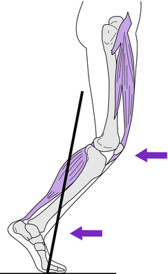
During stance, loading the heel initiates motion at each joint. Prompt response by the muscles provides an eccentric force that limits the arc of joint motion and also serves as a shock-absorbing mechanism. Recent investigations of eccentric muscle function, using portable ultrasound sensors (taped over the target muscle) have differentiated muscle fascicles from tendon during walking and jumping. These findings have redefined eccentric muscle action, which commonly is defined as a lengthening contraction. However, an ultrasound display of the muscle fascicles showed no increase in length.9 The gain in length was stretch of the tendon while the muscle exerted an isometric contraction to stabilize the joint; muscles only shorten or maintain neutral length.12


Ankle and foot


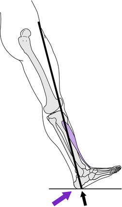
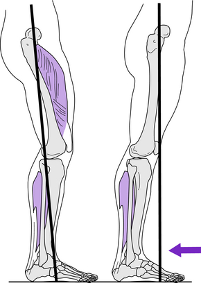
During each stride, the ankle passes through four arcs of motion (Fig. 5-1). At the onset of stance, the ankle is in neutral dorsiflexion, and floor contact is by the heel. Rapid loading of the heel causes the ankle to quickly plantar flex and then return to neutral before forefoot contact. Ultrasound analysis of muscle function at this time identified that the motion was the result of tibialis anterior tendon stretch.6 Release of the stretch force occurred as the heel lever shortened with the advancement of the vector across the heel. Following forefoot contact with the ground, ankle motion reverses to 10 degrees dorsiflexion as the tibia advances over the stationary foot for stance limb progression. Then the ankle plantar flexes 20 degrees during the final phase of stance (preswing). As toe-off starts swing, the foot again dorsiflexes under control of the pretibial muscles. Full elevation of the foot to neutral, however, is not completed until midswing.


The subtalar joint moves into eversion following initial floor contact by the heel. This unlocks the midtarsal joint, allowing it to dorsiflex slightly (arch flattens) following forefoot impact with the floor. Then the subtalar joint progressively inverts and locks the midtarsal joint through late midstance and terminal stance.


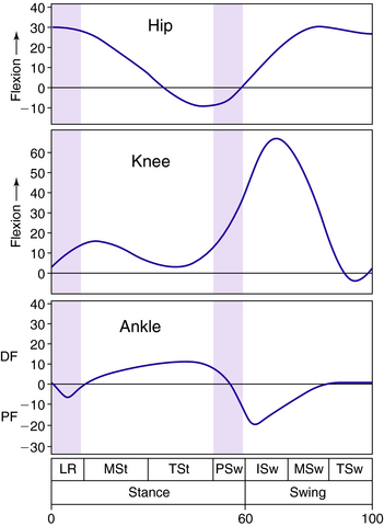
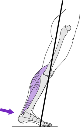
MP joint dorsiflexion is an essential component of heel rise. The foot rolls up over the base of the toes, particularly the great toe, as the trailing limb advances.14


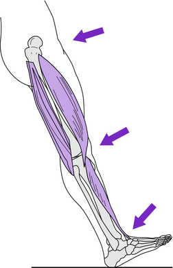
Dorsiflexion control is provided by the pretibial muscles (tibialis anterior, extensor hallucis longus, and extensor digitorum) during the loading response in addition to swing. The soleus and gastrocnemius control the tibia during stance limb progression. During terminal stance the EMG intensity of the gastrocnemius and soleus muscle mass increases rapidly in response to the dorsiflexion moment generated by the advancement of the body mass over the forefoot rocker. This same moment also stretches the tendon and gains 5 degrees of dorsiflexion at the ankle.9 In preswing, the tension of the Achilles tendon is abruptly released by the rapid transfer of body weight to the other limb. This creates a large power burst of plantar flexion by elastic recoil. The muscle mass is inactive (no EMG).9 The “push off” power generated is sufficient to initiate swing.12


Subtalar inversion control is created by the anterior tibialis, posterior tibialis, and soleus. The peroneus brevis and peroneus longus muscles restrain inversion as they produce an eversion force on the lateral side of the foot. Midtarsal restraint of the dorsiflexing forces created by body weight advancement is provided by the intrinsic flexor muscles as well as the subtalar muscles and the long toe flexors. The primary role of the flexor hallucis longus and flexor digitorum longus is to stabilize the MP joint during heel rise.


Knee


Within each gait cycle, the knee alternately flexes and extends both in stance and in swing (see Fig. 5-1). From a position of full extension at initial contact, the knee rapidly flexes 18 degrees during weight acceptance. Ultrasound analysis shows this motion is the result of patellar tendon stretch while the quadriceps muscle is undergoing an isometric contraction.6 This is followed by progressive extension throughout the period of single stance, reaching a final position of 5 degrees flexion. The knee then rapidly flexes to 40 degrees during preswing and continues to 60 degrees in initial swing. From this position, the knee then extends to neutral.


The quadriceps restrains knee flexion in stance and assists extension. All the vasti respond simultaneously. The gluteus maximus through its iliotibial band insertion also contributes to knee extensor stability. Brief and occasional action of the rectus femoris (and less frequently the vastus intermedius) restrains excessive preswing flexion. Knee flexion in swing is aided by the short head of the biceps femoris. Terminal swing knee extension is limited by the hamstring muscle group.


Hip


The major hip motions occur in the plane of progression (see Fig. 5-1). This consists of an arc of extension through stance, reaching 10 degrees hyperextension in terminal stance. A similar arc of flexion occurs from preswing through midswing. The resulting 35-degree flexed posture is maintained in terminal swing and loading response. In the other planes, there are small (4 to 5 degree) arcs of postural accommodation, which are described as pelvic motions.


Hip extensor muscle action begins with the hamstrings in terminal swing and proceeds to the gluteus maximus and adductor magnus during the loading response. Lateral stability of the hip in stance is provided by the gluteus medius–gluteus minimus complex and the tensor fascia lata.


Hip flexion results from serial activation of several muscles: adductor longus (plus rectus femoris), iliacus, sartorius, and gracilis.


Integrated function of the limb


Task I: Weight acceptance


Weight acceptance is the first determinant of the ability to walk. Two objectives determine the events that occur during this task: establishment of a stable limb for weight bearing and minimization of the shock of floor impact. The last phase of swing and the first two stance phases are dedicated to optimum weight acceptance.


Rapid, intense action by the hamstring muscles (semimembranosus, semitendinosus, biceps femoris long head) stops hip flexion and terminates swing. These muscles then reduce their intensity and allow the quadriceps to extend the knee. The continuation of mild hamstring action prevents knee hyperextension from the residual tibial momentum. Pretibial muscle action supports the dorsiflexed foot.


Phase 1—Initial contact: Floor contact by the heel is the critical event (Fig. 5-3). Its purpose is to initiate the heel rocker for progression and shock absorption. The significant postures are ankle dorsiflexion and full knee extension. Initial floor contact by the heel is a forceful event, which begins with 1 cm of free fall between the foot and the ground. The impact registers 50% to 125% body weight during the first 10 to 20 ms of stance (1% to 2% of the gait cycle).17,19 The heel responds to the impact by initiating small arcs of ankle plantar flexion and subtalar inversion. Anterior tibialis control of the foot determines heel rocker effectiveness.


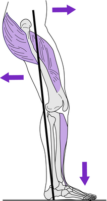
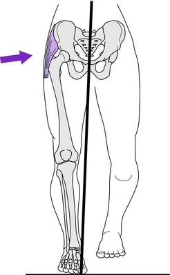
For both shock absorption and progression, the heel rocker drives the foot toward the floor as the limb is loaded. Response of the pretibial muscles to decelerate the dropping foot pulls the tibia forward. This places the vector behind the knee, leading to rapid knee flexion for shock absorption. Prompt quadriceps response opposes the vector’s flexor moment to preserve knee stability and absorb the shock of the initial floor impact. Knee extensor stability is aided by the femoral stability gained from the adductor magnus and gluteus maximus. Prompt relaxation of the hamstring muscles avoids an unnecessary flexor force.


At the hip, there is a rapid response by the abductor muscle group to stabilize the pelvis, which lost its contralateral support with the transfer of body weight to the forward limb.


Task II: Stance limb progression


The basic function is advancement of the limb (and body) over the supporting foot. This is the second determinant of the ability to walk. Two phases of single-limb support are involved as the means of progression differ.


Phase 3—Midstance: The critical event is ankle dorsiflexion for progression of the stance limb over a stationary, flat foot (Fig. 5-5). As momentum from the contralateral swing limb moves the vector along the foot, the soleus (quickly assisted by the gastrocnemius) modulates the tibial advancement so the lower leg proceeds less rapidly than the femur. This provides passive extension of the hip and knee for weight-bearing stability. As a result, the hip extensor and quadriceps muscles rapidly relax and stability of the hip and knee become dependent on the strength of the plantar flexor muscles.


Task III: Limb advancement


The ability to lift the foot is the third determinant of walking ability. Flexing the limb for floor clearance and swing advancement begins in the terminal double-support period of stance. Because the purpose is limb advancement rather than weight bearing, the phase has been titled preswing. The other actions occur throughout swing.


Following floor contact by the other foot, body weight is rapidly transferred to that limb to catch the forward fall. The equally abrupt unloading of the trailing limb initiates a series of actions commonly called push-off. A rapid arc of ankle plantar flexion to 20 degrees is accompanied by passive knee flexion to 40 degrees, increased toe dorsiflexion, and release of the extended hip. The initial force is a large burst of plantar flexion power. Because there is no corresponding EMG, the source of the power is attributed to elastic energy generated by the abrupt release of the previously tense soleus and gastrocnemius muscles: push-off positions the limb for swing and initiates the action, allowing several small forces to be effective. As the limb’s trailing posture reduces the foot’s floor contact to the anterior margins of the metatarsal heads and the toes (fourth rocker), there is no stabilizing force, so the foot and the leg are free to roll forward. This is accelerated by the rapid ankle plantar flexion stimulated by the release of the tension stored in the eccentrically stretched soleus and gastrocnemius. Passive knee flexion is initiated. Unloading the limb also releases the tension in the hip flexors. This force combined with adductor longus action initiates early hip flexion and assists knee flexion.


Phase 6—Initial swing: The critical event is knee flexion sufficient for the toe to clear the floor as the thigh advances. This involves total limb flexion (Fig. 5-9). Hip flexion may be a passive continuation of the preswing events or result from direct action by the iliacus, sartorius, and gracilis. Attainment of full knee flexion largely depends on the imbalance between the forward momentum of the femur generated by hip flexion and inertia of the tibia. Active assistance also is provided by the biceps femoris, short head. Brisk activation of the pretibial muscles initiates ankle dorsiflexion, but the arc is incomplete in initial swing.
< div class='tao-gold-member'>

Only gold members can continue reading. Log In or Register to continue

Stay updated, free articles. Join our Telegram channel

Jul 12, 2016 | Posted by in ORTHOPEDIC | Comments Off on Normal and pathologic gait

Full access? Get Clinical Tree

Get Clinical Tree app for offline access