CHAPTER 10 Nonoperative rehabilitation for traumatic and atraumatic glenohumeral instability


Introduction
A well-designed and appropriate rehabilitation program plays a vital role in the successful outcome following a shoulder instability episode. Shoulder instability is a common pathology often seen in the orthopaedic and sports medicine setting. The glenohumeral joint allows tremendous amounts of joint mobility, thus making it inherently unstable and the most frequently dislocated joint in the body.1 Because of the joint’s poor osseous congruency and capsular laxity, it greatly relies on the dynamic stabilizers and neuromuscular system to provide functional stability.2 Therefore, differentiation between normal translation and pathologic instability is often difficult to determine. A wide range of shoulder instabilities exist, ranging from subtle subluxations (as seen in overhead athletes) to gross instability. Often the success of the rehabilitation program is based on accurate recognition of the specific type of instability present and the treatment program designed to address it.
Rehabilitation factors
There are nine key factors that should be considered when designing a rehabilitation program for a patient with an unstable shoulder (Table 10-1). We briefly discuss these factors and their significance to the rehabilitation program.
Table 10-1 Nine Key Factors to Consider in the Rehabilitation of the Unstable Shoulder
Onset of pathology
Conversely, a patient presenting with atraumatic instability often presents with a history of repetitive injuries and symptomatic complaints. Often the patient does not complain of a single instability episode but rather a feeling of shoulder laxity or an inability to perform specific tasks. Rehabilitation for this patient should focus on early proprioception training, dynamic stabilization drills, neuromuscular control, scapular stabilization exercises, core training, and muscle strengthening exercises to enhance dynamic stability due to the unique characteristic of excessive capsular laxity and capsular redundancy in this type of patient.
Degree of instability
The second factor is the degree of instability present in the patient and its effect on their function. There are varying degrees of shoulder instability, such as a subtle subluxation to gross (uncontrollable) instability. The term subluxation refers to the complete separation of the articular surfaces with spontaneous reduction. Conversely, a dislocation is a complete separation of the articular surfaces and requires a specific movement or manual reduction to relocate the joint, which will result in underlying capsular tissue trauma. Thus, with shoulder dislocations the degree of trauma to the glenohumeral joint’s soft tissue is much more extensive. Speer et al3 have reported that in order for a shoulder dislocation to occur, both a Bankart lesion and soft tissue trauma must be present on both sides of the glenohumeral joint capsule. Thus, in the situation of an acute traumatic dislocation, the anterior capsule may be avulsed off the glenoid (Bankart lesion), and the posterior capsule may be stretched, allowing the humeral head to dislocate. This has been referred to as the “circle stability concept” as described by Warren et al.4 The rate of progression will vary based on the degree of instability and persistence of symptoms. For example, a patient with mild subluxations and muscle guarding may initially tolerate strengthening exercises and neuromuscular control drills more than a patient with a significant amount of muscular guarding.
Frequency
The next factor to influence the rehabilitation program is the frequency of dislocation or subluxation. The primary traumatic dislocation is most often treated conservatively with immobilization in a sling and early controlled passive range of motion (PROM) exercises, especially with first-time dislocations. The incidence of recurrent dislocation ranges from 17% to 96% with a mean of 67% in patient populations between the ages of 21 to 30 years old.1,5–15 Therefore, the rehabilitation program should progress cautiously in young athletic individuals. It should be noted that Hovelius et al8,16–18 have demonstrated that the rate of recurrent dislocations is based on the patient’s age and is not affected by the length of postinjury immobilization. Individuals between the ages of 19 and 29 years are the most likely to experience multiple episodes of instability. Hovelius et al8,16–18 noted patients in their twenties exhibited a recurrence rate of 60%, whereas patients in their thirties to forties had less than a 20% recurrence rate. In adolescents, the recurrence rate is as high as 92%19 and 100% with an open physes.20
Direction of instability
The fourth factor is the direction of instability present. The three most common forms include anterior, posterior, and multidirectional. Anterior instability is the most common traumatic type of instability seen in the general orthopaedic population. It has been reported that this type of instability represents approximately 95% of all traumatic shoulder instabilities.12 However, the incidence of posterior instabilities appears to be dependent on the patient population. For example, in professional or collegiate football, the incidence of posterior shoulder instability appears higher than the general population. This is especially true in linemen because of the pushing methods employed during the blocking motion. Mair et al21 reported on nine athletes with posterior instability in which eight of nine were linemen, and seven were offensive linemen. Often, these patients require surgery as Mair et al21 also reported 75% required surgical stabilization. Kaplan et al22 reported in a study of collegiate football players with shoulder instability that 78% required surgical stabilization.
Following a traumatic event in which the humeral head is forced into extremes of abduction and external rotation, or horizontal abduction, the glenolabral complex and capsule may become detached from the glenoid rim resulting in anterior instability. This type of detachment is referred to as a Bankart lesion, of which there are numerous types. Baker et al23 have identified four types of Bankart lesions based on the size and the degree of tissue involvement. Conversely, rarely will a patient with atraumatic instability and capsular redundancy dislocate their shoulder. It is the author’s opinion that they are more likely to repeatedly sublux the joint without complete separation of the humerus from the glenoid rim. Capsular avulsions can occur on the glenoid side (Bankart lesion) or on the humeral head side referred to as a humeral avulsion of the inferior glenohumeral ligament (HAGL) lesion.24–26
Posterior instability occurs less frequently, only accounting for less than 5% of traumatic shoulder dislocations.27–28 This type of instability is often seen following a traumatic event such as falling onto an outstretched hand or from a pushing mechanism. Furthermore, posterior instability may develop secondary to swinging a bat or golf club during the follow-through phase. However, patients with significant atraumatic laxity may complain of posterior instability especially with shoulder elevation, horizontal adduction, and excessive internal rotation because of the strain placed on the posterior capsule in these positions.
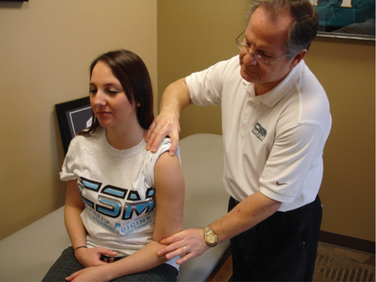
Multidirectional instability (MDI) can be identified as shoulder instability in more than one plane of motion. Patients with MDI have a congenital predisposition and exhibit ligamentous laxity because of excessive collagen elasticity of the capsule. Furthermore, Rodeo et al29 reported that this type of patient exhibits a greater concentration of elastin compared to collagen and also smaller diameter collagen fibrils. The authors consider an inferior displacement of greater than 8 mm to 10 mm during the sulcus maneuver (Fig. 10-1) with the arm adducted to the side as significant hypermobility, thus suggesting significant congenital laxity.2
Because of the atraumatic mechanism and lack of acute tissue damage, ROM is often normal to excessive. Patients with recurrent shoulder instability due to MDI generally have weakness in the rotator cuff, deltoid, and scapular stabilizers with poor dynamic stabilization and inadequate static stabilizers. Initially, the focus is on maximizing dynamic stability, scapula positioning, proprioception, and improving neuromuscular control in mid-ROM. Also, rehabilitation should focus on improving the efficiency and effectiveness of glenohumeral joint force couples through cocontraction exercises, rhythmic stabilization, and neuromuscular control drills. Isotonic strengthening exercises for the rotator cuff, deltoid, and scapular muscles also are emphasized to enhance dynamic stability. Morris et al30 reported the electromyogram (EMG) activity of the rotator cuff and deltoid muscle in MDI and asymptomatic subjects. The authors noted the most significant difference was in the deltoid muscles compared with the rotator cuff muscles in their groups.
Premorbid status of tissue
The fifth factor involves considering other tissues that may have been affected and the premorbid status of the tissue. Disruption of the anterior capsulolabral complex from the glenoid commonly occurs during a traumatic injury, resulting in an anterior Bankart lesion. Often osseous lesions may be present such as a concomitant Hill-Sachs lesion caused by an impaction of the posterolateral aspect of the humeral head as it compresses against the anterior glenoid rim during relocation. This has been reported in up to 80% of dislocations.31–33 Conversely, a reverse Hill-Sachs lesion may be present on the anterior aspect of the humeral head because of a posterior dislocation.34 Occasionally, a bone bruise may be present in individuals who have sustained a shoulder dislocation, thereby restricting upper extremity weight-bearing activities early on in the rehabilitation process. In rare cases of extreme trauma, the brachial plexus may become involved as well.35 Burkhart et al36 reported some patients exhibited a boney defect or inverted pear-shaped glenoid that resulted in recurrent instability if not accurately identified or properly treated. Other common injuries in the unstable shoulder may involve the superior labrum (SLAP lesion) such as a type V SLAP lesion characterized by a Bankart lesion of the anterior capsule extending into the anterior superior labrum.37 Injuries to the rotator cuff also may be observed and significantly affect the rehabilitation progression and long-term function of the patient. These concomitant lesions will affect the rehabilitation significantly in order to protect the healing tissue.
Neuromuscular control
The sixth factor to consider is the patient’s level of neuromuscular control, particularly at end range. Neuromuscular control may be defined as the efferent, or motor output in reaction to an afferent, or sensory input.2,10 The afferent input is the ability to detect the glenohumeral joint position and motion in space with resultant efferent response by the dynamic stabilizers as they blend with the joint capsule to assist in stabilization of the humeral head. Injury with resultant insufficient neuromuscular control could result in deleterious effects to the patient. As a result, the humeral head may not center itself within the glenoid, thereby compromising the surrounding static stabilizers. The patient with poor neuromuscular control may exhibit excessive humeral head migration with the potential for injury, an inflammatory response, and reflexive inhibition of the dynamic stabilizers.
Several authors have reported that neuromuscular control of the glenohumeral joint may be negatively affected by joint instability. Lephart et al10 compared the ability to detect passive motion and the ability to reproduce joint positions in normal, unstable, and surgically repaired shoulders. The authors reported a significant decrease in proprioception and kinesthesia in the shoulders with instability when compared with both normal shoulders and shoulders undergoing surgical stabilization procedures. Smith et al38 reported a significant decrease in proprioception following a shoulder dislocation. Blasier et al39 reported that individuals with significant capsular laxity exhibited a decrease in proprioception compared with patients with normal laxity. Zuckerman et al40 noted that proprioception is affected by the patient’s age, with older subjects exhibiting diminished proprioception as compared with a comparably younger population. Thus, the patient presenting with traumatic or acquired instability may present with poor proprioception and neuromuscular control.
Arm dominance
The seventh factor to consider in the nonoperative rehabilitation of the unstable shoulder is the arm dominance and the desired activity level of the patient. If the patient frequently performs an overhead motion or sporting activities such as a tennis, volleyball, or a throwing sport, then the rehabilitation program should include sport-specific dynamic stabilization exercises, neuromuscular control drills, and plyometric exercises in the overhead position once full, pain-free ROM and adequate strength have been achieved. Patients whose functional demands involve below shoulder level activities will follow a progressive exercise program to return full ROM and strength. The success rates of patients returning to overhead sports after a traumatic dislocation of their dominant arm are extremely low.41 Arm dominance can also significantly influence the successful outcome. The recurrence rates of instabilities vary based on age, activity level, and arm dominance. In athletes involved in collision sports, the recurrence rates have been reported between 86% to 94%.6,42–44
Patient age
The next factor is the age of the patient. Younger patients (ages 17 to 24) tend to exhibit a different lesion than older patients (older than 40 years of age). Younger patients usually exhibit a Bankart lesion, and some may exhibit a HAGL lesion; conversely, older patients may exhibit an anterior labral periosteal sleeve avulsion (ALPSA) lesion.45
Rehabilitation guidelines
Traumatic shoulder instability
The following pink-highlighted text outlines the nonoperative rehabilitation protocol for traumatic dislocation of the shoulder.* The program will vary in length for each individual depending on the following factors:
Phase I—acute motion phase
NOTE: Do not stretch injured capsule
NOTE: Motion is performed in nonpainful arc of motion only.
Do not push into ER or horizontal abduction with anterior instability.
Avoid excessive IR or horizontal adduction with posterior instability.
NOTE: Electrical muscle stimulation may be used to ER during isometrics
Phase II—intermediate phase
Criteria to progress to phase II:
NOTE: Electrical muscle stimulation may be used to ER during exercises
Phase III—advanced strengthening phase
Criteria to progress to phase III:
Phase IV—return to activity phase
Criteria to progress to phase IV:
Related posts:
 Recurrent instability due to capsular deficiency
Open treatment of anterior instability—surgical technique
Arthroscopic treatment of multidirectional instability—surgical technique
Recognition and management of combined instability and rotator cuff tears
Arthroscopic treatment of posterior instability—surgical technique
Clinical anatomy and biomechanics of the glenohumeral joint (including stabilizers)
Recurrent instability due to capsular deficiency
Open treatment of anterior instability—surgical technique
Arthroscopic treatment of multidirectional instability—surgical technique
Recognition and management of combined instability and rotator cuff tears
Arthroscopic treatment of posterior instability—surgical technique
Clinical anatomy and biomechanics of the glenohumeral joint (including stabilizers)
Stay updated, free articles. Join our Telegram channel

Full access? Get Clinical Tree


